8QYH | pdb_00008qyh

Zorya anti-bacteriophage defense system ZorAB ZorA E86A_E89A, Calcium binding site mutation


Experimental Data Snapshot

  • Method: ELECTRON MICROSCOPY
  • Resolution: 2.40 Å
  • Aggregation State: PARTICLE 
  • Reconstruction Method: SINGLE PARTICLE 

wwPDB Validation   3D Report Full Report


This is version 1.2 of the entry. See complete history


Literature

Structure and mechanism of the Zorya anti-phage defence system.

Hu, H.Popp, P.F.Hughes, T.C.D.Roa-Eguiara, A.Rutbeek, N.R.Martin, F.J.O.Hendriks, I.A.Payne, L.J.Yan, Y.Humolli, D.Klein-Sousa, V.Songailiene, I.Wang, Y.Nielsen, M.L.Berry, R.M.Harms, A.Erhardt, M.Jackson, S.A.Taylor, N.M.I.

(2025) Nature 639: 1093-1101

  • DOI: https://doi.org/10.1038/s41586-024-08493-8
  • Primary Citation of Related Structures:  
    8QY7, 8QYC, 8QYD, 8QYH, 8QYK, 8QYY, 8R68

  • PubMed Abstract: 

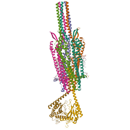
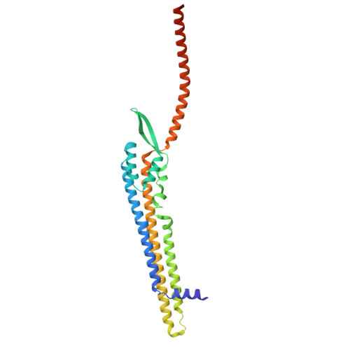
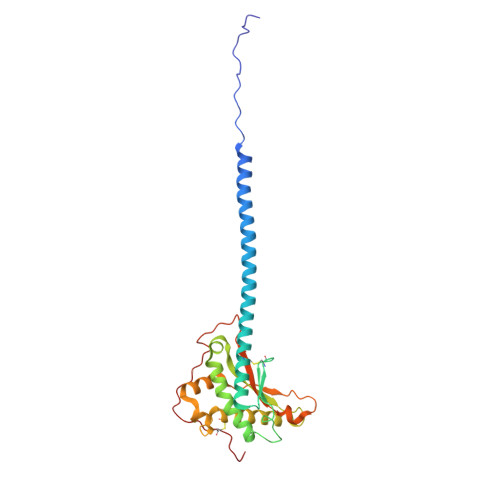
    Zorya is a recently identified and widely distributed bacterial immune system that protects bacteria from viral (phage) infections. Three Zorya subtypes have been discovered, each containing predicted membrane-embedded ZorAB complexes paired with soluble subunits that differ among Zorya subtypes, notably ZorC and ZorD in type I Zorya systems 1,2 . Here, we investigate the molecular basis of Zorya defense using cryo-electron microscopy, mutagenesis, fluorescence microscopy, proteomics, and functional studies. We present cryo-EM structures of ZorAB and show that it shares stoichiometry and features of other 5:2 inner membrane ion-driven rotary motors. The ZorA 5 B 2 complex contains a dimeric ZorB peptidoglycan binding domain and a pentameric α-helical coiled-coil tail made of ZorA that projects approximately 70 nm into the cytoplasm. We also characterize the structure and function of the soluble Zorya components, ZorC and ZorD, finding that they harbour DNA binding and nuclease activity, respectively. Comprehensive functional and mutational analyses demonstrate that all Zorya components work in concert to protect bacterial cells against invading phages. We provide evidence that ZorAB operates as a proton-driven motor that becomes activated upon sensing of phage invasion. Subsequently, ZorAB transfers the phage invasion signal through the ZorA cytoplasmic tail to recruit and activate the soluble ZorC and ZorD effectors, which facilitate degradation of the phage DNA. In summary, our study elucidates the foundational mechanisms of Zorya function as an anti-phage defense system.


  • Organizational Affiliation

    Structural Biology of Molecular Machines Group, Protein Structure & Function Program, Novo Nordisk Foundation Center for Protein Research, Faculty of Health and Medical Sciences, University of Copenhagen, Copenhagen, Denmark. haidai.hu@cpr.ku.dk.


Macromolecules
Find similar proteins by:  (by identity cutoff)  |  3D Structure
Entity ID: 1
MoleculeChains Sequence LengthOrganismDetailsImage
Anti-phage defense ZorAB system ZorA
A, B, C, D, E
280Escherichia coliMutation(s): 0 
Gene Names: zorA
Membrane Entity: Yes 
UniProt
Find proteins for A7ZI10 (Escherichia coli O139:H28 (strain E24377A / ETEC))
Explore A7ZI10 
Go to UniProtKB:  A7ZI10
Entity Groups  
Sequence Clusters30% Identity50% Identity70% Identity90% Identity95% Identity100% Identity
UniProt GroupA7ZI10
Sequence Annotations
Expand
  • Reference Sequence
Find similar proteins by:  (by identity cutoff)  |  3D Structure
Entity ID: 2
MoleculeChains Sequence LengthOrganismDetailsImage
Membrane protein
F, G
246Escherichia coliMutation(s): 0 
Gene Names: AW119_26350
Membrane Entity: Yes 
UniProt
Find proteins for A7ZI09 (Escherichia coli O139:H28 (strain E24377A / ETEC))
Explore A7ZI09 
Go to UniProtKB:  A7ZI09
Entity Groups  
Sequence Clusters30% Identity50% Identity70% Identity90% Identity95% Identity100% Identity
UniProt GroupA7ZI09
Sequence Annotations
Expand
  • Reference Sequence
Small Molecules
Ligands 2 Unique
IDChains Name / Formula / InChI Key2D Diagram3D Interactions
CDL (Subject of Investigation/LOI)
Query on CDL

Download Ideal Coordinates CCD File 
H [auth A],
J [auth B],
L [auth C],
N [auth D],
P [auth E]
CARDIOLIPIN
C81 H156 O17 P2
XVTUQDWPJJBEHJ-KZCWQMDCSA-L
PEE
Query on PEE

Download Ideal Coordinates CCD File 
I [auth A],
K [auth B],
M [auth C],
O [auth D],
Q [auth E]
1,2-dioleoyl-sn-glycero-3-phosphoethanolamine
C41 H78 N O8 P
MWRBNPKJOOWZPW-NYVOMTAGSA-N
Experimental Data & Validation

Experimental Data

  • Method: ELECTRON MICROSCOPY
  • Resolution: 2.40 Å
  • Aggregation State: PARTICLE 
  • Reconstruction Method: SINGLE PARTICLE 

Structure Validation

View Full Validation Report


Entry History & Funding Information

Deposition Data


Funding OrganizationLocationGrant Number
Novo Nordisk FoundationDenmarkNNF17OC0031006
Novo Nordisk FoundationDenmarkNNF21OC0071948
LundbeckfondenDenmarkR347-2020-2429

Revision History  (Full details and data files)

  • Version 1.0: 2024-11-06
    Type: Initial release
  • Version 1.1: 2025-02-12
    Changes: Data collection, Database references
  • Version 1.2: 2025-04-09
    Changes: Data collection, Database references