Dual Modes of Modification of Hepatitis A Virus 3C Protease by a Serine-derived beta-Lactone: Selective Crystallization and Formation of a Functional Catalytic Triad in the Active Site
Yin, J., Bergmann, E.M., Cherney, M.M., Lall, M.S., Jain, R.P., Vederas, J.C., James, M.N.G.(2005) J Mol Biology 354: 854-871
- PubMed: 16288920
- DOI: https://doi.org/10.1016/j.jmb.2005.09.074
- Primary Citation of Related Structures:
2A4O, 2CXV - PubMed Abstract:
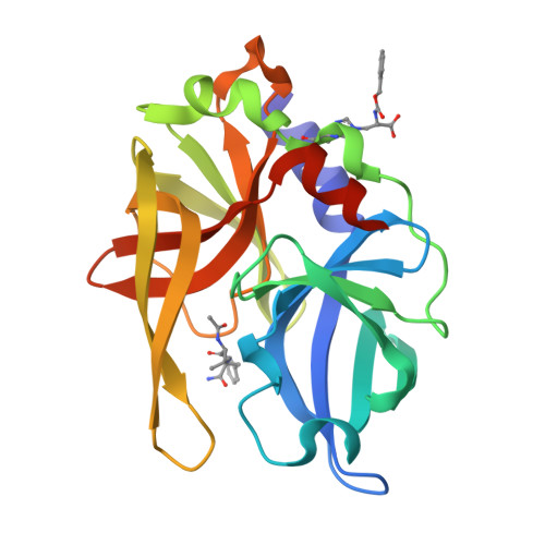
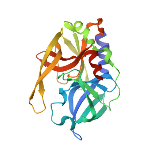
Hepatitis A virus (HAV) 3C proteinase is a member of the picornain cysteine proteases responsible for the processing of the viral polyprotein, a function essential for viral maturation and infectivity. This and its structural similarity to other 3C and 3C-like proteases make it an attractive target for the development of antiviral drugs. Previous solution NMR studies have shown that a Cys24Ser (C24S) variant of HAV 3C protein, which displays catalytic properties indistinguishable from the native enzyme, is irreversibly inactivated by N-benzyloxycarbonyl-l-serine-beta-lactone (1a) through alkylation of the sulfur atom at the active site Cys172. However, crystallization of an enzyme-inhibitor adduct from the reaction mixture followed by X-ray structural analysis shows only covalent modification of the epsilon2-nitrogen of the surface His102 by the beta-lactone with no reaction at Cys172. Re-examination of the heteronuclear multiple quantum coherence (HMQC) NMR spectra of the enzyme-inhibitor mixture indicates that dual modes of single covalent modification occur with a >/=3:1 ratio of S-alkylation of Cys172 to N-alkylation of His102. The latter product crystallizes readily, probably due to the interaction between the phenyl ring of the N-benzyloxycarbonyl (N-Cbz) moiety and a hydrophobic pocket of a neighboring protein molecule in the crystal. Furthermore, significant structural changes are observed in the active site of the 3C protease, which lead to the formation of a functional catalytic triad with Asp84 accepting one hydrogen bond from His44. Although the 3C protease modified at Cys172 is catalytically inactive, the singly modified His102 N(epsilon2)-alkylated protein displays a significant level of enzymatic activity, which can be further modified/inhibited by N-iodoacetyl-valine-phenylalanine-amide (IVF) (in solution and in crystal) or excessive amount of the same beta-lactone inhibitor (in solution). The success of soaking IVF into HAV 3C-1a crystals demonstrates the usefulness of this new crystal form in the study of enzyme-inhibitor interactions in the proteolytic active site.
Organizational Affiliation:
CIHR Group in Protein Structure and Function, Department of Biochemistry, University of Alberta, Edmonton, Alberta, Canada T6G 2H7.