6PVC | pdb_00006pvc

Structure of human MAIT A-F7 TCR in complex with human MR1-DB28


Experimental Data Snapshot

  • Method: X-RAY DIFFRACTION
  • Resolution: 1.96 Å
  • R-Value Free: 
    0.212 (Depositor), 0.210 (DCC) 
  • R-Value Work: 
    0.169 (Depositor), 0.170 (DCC) 
  • R-Value Observed: 
    0.171 (Depositor) 

Starting Model: experimental
View more details

wwPDB Validation   3D Report Full Report


Ligand Structure Quality Assessment 

Created with Raphaël 2.3.0Worse 01 BetterLigand structure goodness of fit to experimental dataBest fitted P1JClick on this verticalbar to view details

This is version 1.5 of the entry. See complete history


Literature

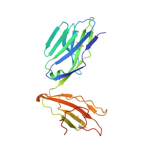
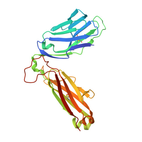
Ligand-dependent downregulation of MR1 cell surface expression.

Salio, M.Awad, W.Veerapen, N.Gonzalez-Lopez, C.Kulicke, C.Waithe, D.Martens, A.W.J.Lewinsohn, D.M.Hobrath, J.V.Cox, L.R.Rossjohn, J.Besra, G.S.Cerundolo, V.

(2020) Proc Natl Acad Sci U S A 117: 10465-10475

  • DOI: https://doi.org/10.1073/pnas.2003136117
  • Primary Citation of Related Structures:  
    6PVC, 6PVD

  • PubMed Abstract: 

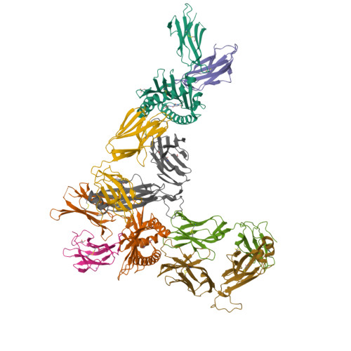
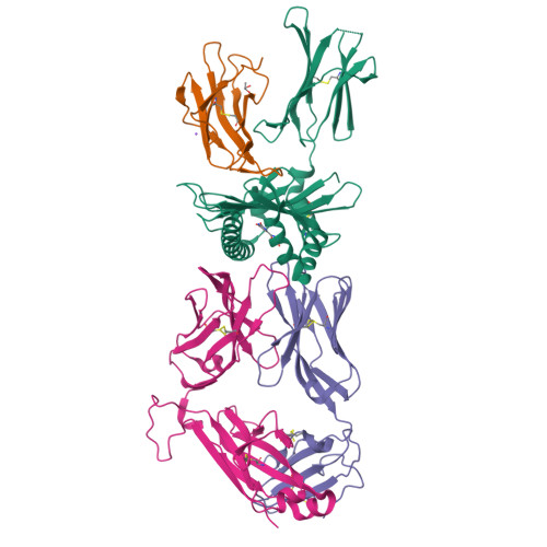
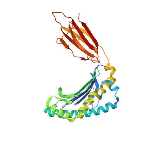
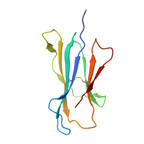
    The antigen-presenting molecule MR1 presents riboflavin-based metabolites to Mucosal-Associated Invariant T (MAIT) cells. While MR1 egress to the cell surface is ligand-dependent, the ability of small-molecule ligands to impact on MR1 cellular trafficking remains unknown. Arising from an in silico screen of the MR1 ligand-binding pocket, we identify one ligand, 3-([2,6-dioxo-1,2,3,6-tetrahydropyrimidin-4-yl]formamido)propanoic acid, DB28, as well as an analog, methyl 3-([2,6-dioxo-1,2,3,6-tetrahydropyrimidin-4-yl]formamido)propanoate, NV18.1, that down-regulate MR1 from the cell surface and retain MR1 molecules in the endoplasmic reticulum (ER) in an immature form. DB28 and NV18.1 compete with the known MR1 ligands, 5-OP-RU and acetyl-6-FP, for MR1 binding and inhibit MR1-dependent MAIT cell activation. Crystal structures of the MAIT T cell receptor (TCR) complexed with MR1-DB28 and MR1-NV18.1, show that these two ligands reside within the A'-pocket of MR1. Neither ligand forms a Schiff base with MR1 molecules; both are nevertheless sequestered by a network of hydrophobic and polar contacts. Accordingly, we define a class of compounds that inhibits MR1 cellular trafficking.


  • Organizational Affiliation

    MRC Human Immunology Unit, MRC Weatherall Institute of Molecular Medicine, University of Oxford, Oxford OX3 9DS, United Kingdom; mariolina.salio@imm.ox.ac.uk g.besra@bham.ac.uk.


Macromolecules
Find similar proteins by:  (by identity cutoff)  |  3D Structure
Entity ID: 1
MoleculeChains Sequence LengthOrganismDetailsImage
Major histocompatibility complex class I-related gene protein
A, C
271Homo sapiensMutation(s): 0 
Gene Names: MR1
UniProt & NIH Common Fund Data Resources
Find proteins for Q95460 (Homo sapiens)
Explore Q95460 
Go to UniProtKB:  Q95460
PHAROS:  Q95460
GTEx:  ENSG00000153029 
Entity Groups  
Sequence Clusters30% Identity50% Identity70% Identity90% Identity95% Identity100% Identity
UniProt GroupQ95460
Sequence Annotations
Expand
  • Reference Sequence
Find similar proteins by:  (by identity cutoff)  |  3D Structure
Entity ID: 2
MoleculeChains Sequence LengthOrganismDetailsImage
Beta-2-microglobulin
B, D
100Homo sapiensMutation(s): 0 
Gene Names: B2MCDABP0092HDCMA22P
UniProt & NIH Common Fund Data Resources
Find proteins for P61769 (Homo sapiens)
Explore P61769 
Go to UniProtKB:  P61769
PHAROS:  P61769
GTEx:  ENSG00000166710 
Entity Groups  
Sequence Clusters30% Identity50% Identity70% Identity90% Identity95% Identity100% Identity
UniProt GroupP61769
Sequence Annotations
Expand
  • Reference Sequence
Find similar proteins by:  (by identity cutoff)  |  3D Structure
Entity ID: 3
MoleculeChains Sequence LengthOrganismDetailsImage
Human TCR alpha chain
E, G
204Homo sapiensMutation(s): 0 
Entity Groups  
Sequence Clusters30% Identity50% Identity70% Identity90% Identity95% Identity100% Identity
Sequence Annotations
Expand
  • Reference Sequence
Find similar proteins by:  (by identity cutoff)  |  3D Structure
Entity ID: 4
MoleculeChains Sequence LengthOrganismDetailsImage
Human TCR beta chain
F, H
246Homo sapiensMutation(s): 0 
Entity Groups  
Sequence Clusters30% Identity50% Identity70% Identity90% Identity95% Identity100% Identity
Sequence Annotations
Expand
  • Reference Sequence
Small Molecules
Ligands 4 Unique
IDChains Name / Formula / InChI Key2D Diagram3D Interactions
P1J (Subject of Investigation/LOI)
Query on P1J

Download Ideal Coordinates CCD File 
J [auth A],
K [auth C]
N-(2,6-dioxo-1,2,3,6-tetrahydropyrimidine-4-carbonyl)-beta-alanine
C8 H9 N3 O5
HLVBXSGXJVQJGW-UHFFFAOYSA-N
GOL
Query on GOL

Download Ideal Coordinates CCD File 
I [auth A],
M [auth D]
GLYCEROL
C3 H8 O3
PEDCQBHIVMGVHV-UHFFFAOYSA-N
ACT
Query on ACT

Download Ideal Coordinates CCD File 
N [auth H]ACETATE ION
C2 H3 O2
QTBSBXVTEAMEQO-UHFFFAOYSA-M
NA
Query on NA

Download Ideal Coordinates CCD File 
L [auth D]SODIUM ION
Na
FKNQFGJONOIPTF-UHFFFAOYSA-N
Experimental Data & Validation

Experimental Data

  • Method: X-RAY DIFFRACTION
  • Resolution: 1.96 Å
  • R-Value Free:  0.212 (Depositor), 0.210 (DCC) 
  • R-Value Work:  0.169 (Depositor), 0.170 (DCC) 
  • R-Value Observed: 0.171 (Depositor) 
Space Group: C 1 2 1
Unit Cell:
Length ( Å )Angle ( ˚ )
a = 214.291α = 90
b = 69.834β = 103.319
c = 141.618γ = 90
Software Package:
Software NamePurpose
PHENIXrefinement
XDSdata reduction
Aimlessdata scaling
PHASERphasing

Structure Validation

View Full Validation Report


Ligand Structure Quality Assessment 

Created with Raphaël 2.3.0Worse 01 BetterLigand structure goodness of fit to experimental dataBest fitted P1JClick on this verticalbar to view details

Entry History & Funding Information

Deposition Data


Funding OrganizationLocationGrant Number
Australian Research Council (ARC)Australia--

Revision History  (Full details and data files)

  • Version 1.0: 2020-04-01
    Type: Initial release
  • Version 1.1: 2020-05-06
    Changes: Database references
  • Version 1.2: 2020-05-13
    Changes: Database references
  • Version 1.3: 2020-05-27
    Changes: Database references
  • Version 1.4: 2023-10-11
    Changes: Data collection, Database references, Derived calculations, Refinement description
  • Version 1.5: 2024-10-16
    Changes: Structure summary