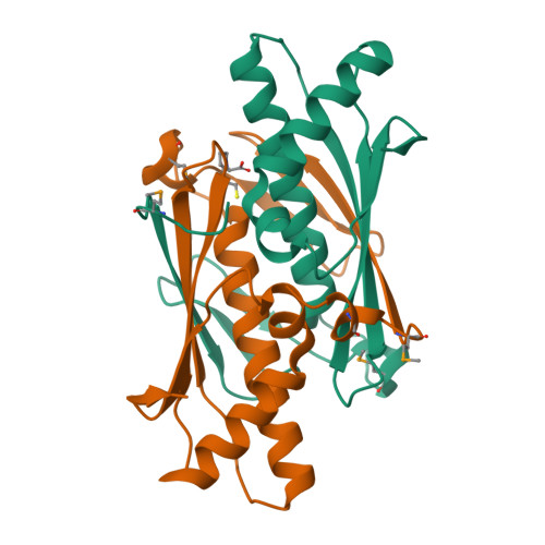
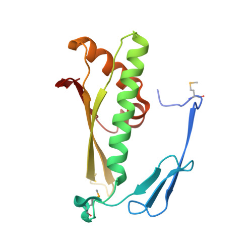
Crystal structure of a putative organic hydroperoxide resistance protein with molecule of captopril bound in one of the active sites from Vibrio cholerae O1 biovar eltor str. N16961
Nocek, B., Maltseva, N., Makowska-Grzyska, N., Kwon, K., Anderson, W., Joachimiak, A., NIAIDTo be published.