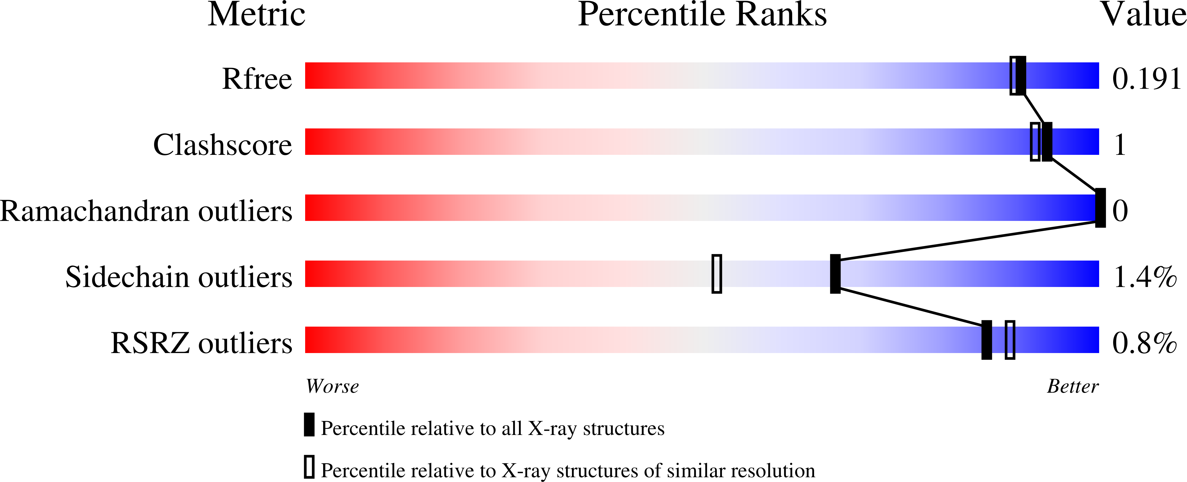
8P4H

Crystal structure of human methionine adenosyltransferase 2A (MAT2A) in complex with SAM and allosteric compound IDEAYA cmpd A


Experimental Data Snapshot

  • Method: X-RAY DIFFRACTION
  • Resolution: 1.71 Å
  • R-Value Free: 0.184 
  • R-Value Work: 0.153 
  • R-Value Observed: 0.154 

Starting Model: experimental
View more details

wwPDB Validation   3D Report Full Report


Ligand Structure Quality Assessment 


This is version 1.2 of the entry. See complete history


Literature

Discovery of novel methionine adenosyltransferase 2A (MAT2A) allosteric inhibitors by structure-based virtual screening.

Kalliokoski, T.Kettunen, H.Kumpulainen, E.Kettunen, E.Thieulin-Pardo, G.Neumann, L.Thomsen, M.Paul, R.Malyutina, A.Georgiadou, M.

(2023) Bioorg Med Chem Lett 94: 129450-129450

  • DOI: https://doi.org/10.1016/j.bmcl.2023.129450
  • Primary Citation of Related Structures:  
    8P1T, 8P1V, 8P1W, 8P4H

  • PubMed Abstract: 

    Methionine adenosyltransferase 2A (MAT2A) has been indicated as a drug target for oncology indications. Clinical trials with MAT2A inhibitors are currently on-going. Here, a structure-based virtual screening campaign was performed on the commercially available chemical space which yielded two novel MAT2A-inhibitor chemical series. The binding modes of the compounds were confirmed with X-ray crystallography. Both series have acceptable physicochemical properties and show nanomolar activity in the biochemical MAT2A inhibition assay and single-digit micromolar activity in the proliferation assay (MTAP -/- cell line). The identified compounds and the relating structural data could be helpful in related drug discovery projects.


  • Organizational Affiliation

    Orion Pharma, Orionintie 1A, 02101 Espoo, Finland. Electronic address: [email protected].


Macromolecules
Find similar proteins by:  (by identity cutoff)  |  3D Structure
Entity ID: 1
MoleculeChains Sequence LengthOrganismDetailsImage
S-adenosylmethionine synthase isoform type-2
A, B, C, D
412Homo sapiensMutation(s): 0 
Gene Names: MAT2AAMS2MATA2
EC: 2.5.1.6
UniProt & NIH Common Fund Data Resources
Find proteins for P31153 (Homo sapiens)
Explore P31153 
Go to UniProtKB:  P31153
PHAROS:  P31153
GTEx:  ENSG00000168906 
Entity Groups  
Sequence Clusters30% Identity50% Identity70% Identity90% Identity95% Identity100% Identity
UniProt GroupP31153
Sequence Annotations
Expand
  • Reference Sequence
Small Molecules
Ligands 5 Unique
IDChains Name / Formula / InChI Key2D Diagram3D Interactions
SAM
Query on SAM

Download Ideal Coordinates CCD File 
K [auth B],
Q [auth D]
S-ADENOSYLMETHIONINE
C15 H22 N6 O5 S
MEFKEPWMEQBLKI-FCKMPRQPSA-N
UM6 (Subject of Investigation/LOI)
Query on UM6

Download Ideal Coordinates CCD File 
E [auth A],
L [auth B],
O [auth C],
S [auth D]
7-chloranyl-4-[(3R)-3-fluoranylpyrrolidin-1-yl]-1-phenyl-quinazolin-2-one
C18 H15 Cl F N3 O
GPTUSHRRTGGGTD-CYBMUJFWSA-N
GOL
Query on GOL

Download Ideal Coordinates CCD File 
F [auth A]
G [auth A]
H [auth A]
I [auth A]
N [auth B]
F [auth A],
G [auth A],
H [auth A],
I [auth A],
N [auth B],
P [auth C],
T [auth D]
GLYCEROL
C3 H8 O3
PEDCQBHIVMGVHV-UHFFFAOYSA-N
CL
Query on CL

Download Ideal Coordinates CCD File 
M [auth B],
U [auth D]
CHLORIDE ION
Cl
VEXZGXHMUGYJMC-UHFFFAOYSA-M
MG
Query on MG

Download Ideal Coordinates CCD File 
J [auth B],
R [auth D]
MAGNESIUM ION
Mg
JLVVSXFLKOJNIY-UHFFFAOYSA-N
Modified Residues  1 Unique
IDChains TypeFormula2D DiagramParent
CSO
Query on CSO
A, B, C, D
L-PEPTIDE LINKINGC3 H7 N O3 SCYS
Binding Affinity Annotations 
IDSourceBinding Affinity
UM6 BindingDB:  8P4H IC50: min: 36, max: 200 (nM) from 2 assay(s)
Experimental Data & Validation

Experimental Data

  • Method: X-RAY DIFFRACTION
  • Resolution: 1.71 Å
  • R-Value Free: 0.184 
  • R-Value Work: 0.153 
  • R-Value Observed: 0.154 
  • Space Group: P 1 21 1
Unit Cell:
Length ( Å )Angle ( ˚ )
a = 67.898α = 90
b = 106.742β = 93.18
c = 114.996γ = 90
Software Package:
Software NamePurpose
REFMACrefinement
XDSdata reduction
autoPROCdata scaling
PHASERphasing

Structure Validation

View Full Validation Report


Ligand Structure Quality Assessment 


Entry History & Funding Information

Deposition Data


Funding OrganizationLocationGrant Number
Not funded--

Revision History  (Full details and data files)

  • Version 1.0: 2023-08-30
    Type: Initial release
  • Version 1.1: 2023-11-15
    Changes: Data collection
  • Version 1.2: 2024-10-23
    Changes: Structure summary