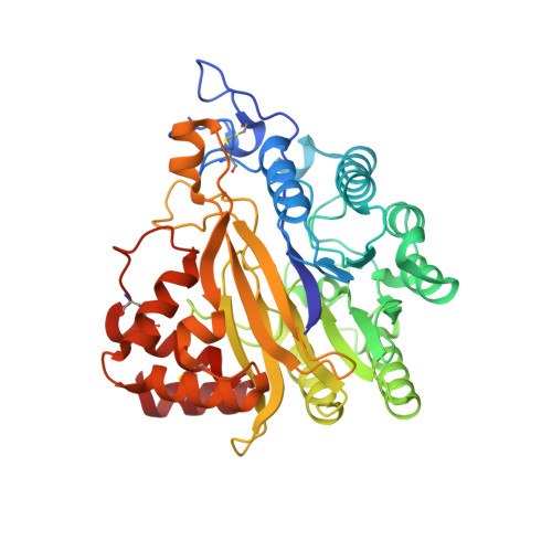
8W6P

Crystal structure of dimeric murine SMPDL3A


Experimental Data Snapshot

  • Method: X-RAY DIFFRACTION
  • Resolution: 1.91 Å
  • R-Value Free: 0.246 
  • R-Value Work: 0.218 
  • R-Value Observed: 0.218 

Starting Model: experimental
View more details

wwPDB Validation   3D Report Full Report


Ligand Structure Quality Assessment 


This is version 1.1 of the entry. See complete history


Literature

SMPDL3A is a cGAMP-degrading enzyme induced by LXR-mediated lipid metabolism to restrict cGAS-STING DNA sensing.

Hou, Y.Wang, Z.Liu, P.Wei, X.Zhang, Z.Fan, S.Zhang, L.Han, F.Song, Y.Chu, L.Zhang, C.

(2023) Immunity 56: 2492-2507.e10

  • DOI: https://doi.org/10.1016/j.immuni.2023.10.001
  • Primary Citation of Related Structures:  
    8W6P, 8W6R

  • PubMed Abstract: 

    Lipid metabolism has been associated with the cyclic guanosine monophosphate (GMP)-AMP synthase (cGAS) stimulator of interferon genes (STING) DNA-sensing pathway, but our understanding of how these signals are integrated into a cohesive immunometabolic program is lacking. Here, we have identified liver X receptor (LXR) agonists as potent inhibitors of STING signaling. We show that stimulation of lipid metabolism by LXR agonists specifically suppressed cyclic GMP-AMP (cGAMP)-STING signaling. Moreover, we developed cyclic dinucleotide-conjugated beads to biochemically isolate host effectors for cGAMP inhibition, and we found that LXR ligands stimulated the expression of sphingomyelin phosphodiesterase acid-like 3A (SMPDL3A), which is a 2'3'-cGAMP-degrading enzyme. Results of crystal structures suggest that cGAMP analog induces dimerization of SMPDL3A, and the dimerization is critical for cGAMP degradation. Additionally, we have provided evidence that SMPDL3A cleaves cGAMP to restrict STING signaling in cell culture and mouse models. Our results reveal SMPDL3A as a cGAMP-specific nuclease and demonstrate a mechanism for how LXR-associated lipid metabolism modulates STING-mediated innate immunity.


  • Organizational Affiliation

    State Key Laboratory of Membrane Biology, School of Pharmaceutical Sciences, Tsinghua University, Beijing 100084, China.


Macromolecules
Find similar proteins by:  (by identity cutoff)  |  3D Structure
Entity ID: 1
MoleculeChains Sequence LengthOrganismDetailsImage
Acid sphingomyelinase-like phosphodiesterase 3a
A, B
413Mus musculusMutation(s): 0 
Gene Names: Smpdl3aAsml3a
EC: 3.1.4 (PDB Primary Data), 3.6.1.15 (UniProt)
UniProt & NIH Common Fund Data Resources
Find proteins for P70158 (Mus musculus)
Explore P70158 
Go to UniProtKB:  P70158
IMPC:  MGI:1931437
Entity Groups  
Sequence Clusters30% Identity50% Identity70% Identity90% Identity95% Identity100% Identity
UniProt GroupP70158
Glycosylation
Glycosylation Sites: 4Go to GlyGen: P70158-1
Sequence Annotations
Expand
  • Reference Sequence
Oligosaccharides

Help

Entity ID: 2
MoleculeChains Length2D Diagram Glycosylation3D Interactions
2-acetamido-2-deoxy-beta-D-glucopyranose-(1-4)-2-acetamido-2-deoxy-beta-D-glucopyranose
C, E
2N-Glycosylation
Glycosylation Resources
GlyTouCan:  G42666HT
GlyCosmos:  G42666HT
GlyGen:  G42666HT
Entity ID: 3
MoleculeChains Length2D Diagram Glycosylation3D Interactions
alpha-L-fucopyranose-(1-6)-2-acetamido-2-deoxy-beta-D-glucopyranose
D
2N-Glycosylation
Glycosylation Resources
GlyTouCan:  G86851RC
GlyCosmos:  G86851RC
GlyGen:  G86851RC
Entity ID: 4
MoleculeChains Length2D Diagram Glycosylation3D Interactions
beta-D-mannopyranose-(1-4)-2-acetamido-2-deoxy-beta-D-glucopyranose-(1-4)-2-acetamido-2-deoxy-beta-D-glucopyranose
F
3N-Glycosylation
Glycosylation Resources
GlyTouCan:  G15407YE
GlyCosmos:  G15407YE
GlyGen:  G15407YE
Small Molecules
Ligands 5 Unique
IDChains Name / Formula / InChI Key2D Diagram3D Interactions
YA4 (Subject of Investigation/LOI)
Query on YA4

Download Ideal Coordinates CCD File 
G [auth A][(2~{R},3~{S},4~{S})-3,4-bis(oxidanyl)oxolan-2-yl]methoxy-sulfanyl-phosphinic acid
C5 H11 O6 P S
HBCHIJUQXWEATM-LMVFSUKVSA-N
NAG (Subject of Investigation/LOI)
Query on NAG

Download Ideal Coordinates CCD File 
H [auth A]
I [auth A]
L [auth B]
N [auth B]
O [auth B]
H [auth A],
I [auth A],
L [auth B],
N [auth B],
O [auth B],
P [auth B]
2-acetamido-2-deoxy-beta-D-glucopyranose
C8 H15 N O6
OVRNDRQMDRJTHS-FMDGEEDCSA-N
FUC (Subject of Investigation/LOI)
Query on FUC

Download Ideal Coordinates CCD File 
M [auth B]alpha-L-fucopyranose
C6 H12 O5
SHZGCJCMOBCMKK-SXUWKVJYSA-N
PO4 (Subject of Investigation/LOI)
Query on PO4

Download Ideal Coordinates CCD File 
Q [auth B]PHOSPHATE ION
O4 P
NBIIXXVUZAFLBC-UHFFFAOYSA-K
ZN (Subject of Investigation/LOI)
Query on ZN

Download Ideal Coordinates CCD File 
J [auth A],
K [auth A],
R [auth B],
S [auth B]
ZINC ION
Zn
PTFCDOFLOPIGGS-UHFFFAOYSA-N
Experimental Data & Validation

Experimental Data

  • Method: X-RAY DIFFRACTION
  • Resolution: 1.91 Å
  • R-Value Free: 0.246 
  • R-Value Work: 0.218 
  • R-Value Observed: 0.218 
  • Space Group: P 41
Unit Cell:
Length ( Å )Angle ( ˚ )
a = 122.225α = 90
b = 122.225β = 90
c = 79.557γ = 90
Software Package:
Software NamePurpose
PHENIXrefinement
PDB_EXTRACTdata extraction
PHENIXmodel building
PHENIXphasing
HKL-2000data reduction
HKL-2000data scaling

Structure Validation

View Full Validation Report


Ligand Structure Quality Assessment 


Entry History & Funding Information

Deposition Data


Funding OrganizationLocationGrant Number
National Natural Science Foundation of China (NSFC)China32070875

Revision History  (Full details and data files)

  • Version 1.0: 2023-12-20
    Type: Initial release
  • Version 1.1: 2024-10-16
    Changes: Structure summary