8UOJ | pdb_00008uoj

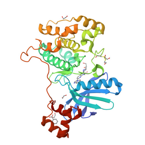
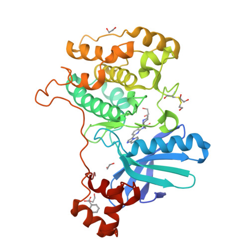
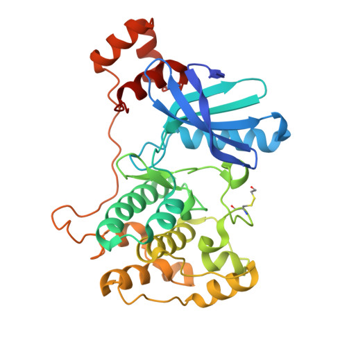
Crystal structure of human NUAK1-MARK3 kinase domain chimera bound with azepane (R)-#50 small molecule inhibitor


Experimental Data Snapshot

  • Method: X-RAY DIFFRACTION
  • Resolution: 1.60 Å
  • R-Value Free: 
    0.186 (Depositor), 0.190 (DCC) 
  • R-Value Work: 
    0.159 (Depositor), 0.163 (DCC) 
  • R-Value Observed: 
    0.160 (Depositor) 

wwPDB Validation   3D Report Full Report


Ligand Structure Quality Assessment 

Created with Raphaël 2.3.0Worse 01 BetterLigand structure goodness of fit to experimental dataBest fitted X5IClick on this verticalbar to view details

This is version 1.2 of the entry. See complete history


Literature

Discovery of UCB9386: A Potent, Selective, and Brain-Penetrant Nuak1 Inhibitor Suitable for In Vivo Pharmacological Studies.

Poullennec, K.G.Jnoff, E.Abendroth, J.Bhuma, N.Calmiano, M.Calmus, L.Cardenas, A.Courade, J.P.Delatour, C.Hall, A.de Haro, T.Delker, S.L.Demaude, T.Gaikwad, N.Ghavate, D.Gholap, A.R.Kierkowicz, M.Le Mestre, R.Van Hijfte, N.Verheijden, S.Vernerova, K.De Wever, V.Waghmode, N.

(2024) J Med Chem 67: 20879-20910

  • DOI: https://doi.org/10.1021/acs.jmedchem.4c01237
  • Primary Citation of Related Structures:  
    8UOJ, 8UOK

  • PubMed Abstract: 

    Nuak1 (NUAK family SnF1-like kinase-1) is a serine-threonine kinase and a member of the AMPK family. Interest in Nuak1 has increased over the years due to the role it plays in several biological processes, from tumor cell invasion and proliferation to Tau stabilization. Nuak1 is expressed in many cancer cell lines and many reports describe this target as an oncogene, the inhibition of which is hypothesized to be valuable for treating various cancer types including glioma. We report here the discovery of Nuak1 inhibitors originating from HTS hit 9 with excellent selectivity and the subsequent medicinal chemistry optimization program, supported by structural information from the first crystal structures of a Nuak1 chimeric protein which provided insights into the binding modes of our compounds. These efforts yielded a nanomolar cell potent, highly selective and brain penetrant Nuak1 inhibitor UCB9386 ( 56 ) suitable for in vivo pharmacological studies for central nervous system (CNS) disorders.


  • Organizational Affiliation

    Chemin du Foriest, UCB, 1420 Braine l'Alleud, Belgium.


Macromolecules
Find similar proteins by:  (by identity cutoff)  |  3D Structure
Entity ID: 1
MoleculeChains Sequence LengthOrganismDetailsImage
MAP/microtubule affinity-regulating kinase 3328Homo sapiensMutation(s): 6 
Gene Names: MARK3CTAK1EMK2
EC: 2.7.11.1
UniProt & NIH Common Fund Data Resources
Find proteins for P27448 (Homo sapiens)
Explore P27448 
Go to UniProtKB:  P27448
PHAROS:  P27448
GTEx:  ENSG00000075413 
Entity Groups  
Sequence Clusters30% Identity50% Identity70% Identity90% Identity95% Identity100% Identity
UniProt GroupP27448
Sequence Annotations
Expand
  • Reference Sequence
Small Molecules
Ligands 4 Unique
IDChains Name / Formula / InChI Key2D Diagram3D Interactions
X5I (Subject of Investigation/LOI)
Query on X5I

Download Ideal Coordinates CCD File 
B [auth A](6M)-4-{[(2R)-azepan-2-yl]methyl}-6-[(4R)-imidazo[1,2-a]pyridin-3-yl]-2H-pyrido[3,2-b][1,4]oxazin-3(4H)-one
C21 H23 N5 O2
YYCHTNBYNGXUNO-OAHLLOKOSA-N
BEZ
Query on BEZ

Download Ideal Coordinates CCD File 
I [auth A]BENZOIC ACID
C7 H6 O2
WPYMKLBDIGXBTP-UHFFFAOYSA-N
EDO
Query on EDO

Download Ideal Coordinates CCD File 
C [auth A],
D [auth A],
E [auth A],
F [auth A],
G [auth A]
1,2-ETHANEDIOL
C2 H6 O2
LYCAIKOWRPUZTN-UHFFFAOYSA-N
CL
Query on CL

Download Ideal Coordinates CCD File 
H [auth A]CHLORIDE ION
Cl
VEXZGXHMUGYJMC-UHFFFAOYSA-M
Modified Residues  1 Unique
IDChains TypeFormula2D DiagramParent
CME
Query on CME
A
L-PEPTIDE LINKINGC5 H11 N O3 S2CYS
Experimental Data & Validation

Experimental Data

  • Method: X-RAY DIFFRACTION
  • Resolution: 1.60 Å
  • R-Value Free:  0.186 (Depositor), 0.190 (DCC) 
  • R-Value Work:  0.159 (Depositor), 0.163 (DCC) 
  • R-Value Observed: 0.160 (Depositor) 
Space Group: I 2 2 2
Unit Cell:
Length ( Å )Angle ( ˚ )
a = 45.35α = 90
b = 115.49β = 90
c = 161.82γ = 90
Software Package:
Software NamePurpose
PHENIXrefinement
XSCALEdata scaling
XDSdata reduction
PHASERphasing

Structure Validation

View Full Validation Report


Ligand Structure Quality Assessment 

Created with Raphaël 2.3.0Worse 01 BetterLigand structure goodness of fit to experimental dataBest fitted X5IClick on this verticalbar to view details

Entry History & Funding Information

Deposition Data


Funding OrganizationLocationGrant Number
Not funded--

Revision History  (Full details and data files)

  • Version 1.0: 2024-11-06
    Type: Initial release
  • Version 1.1: 2024-12-11
    Changes: Database references
  • Version 1.2: 2024-12-25
    Changes: Database references