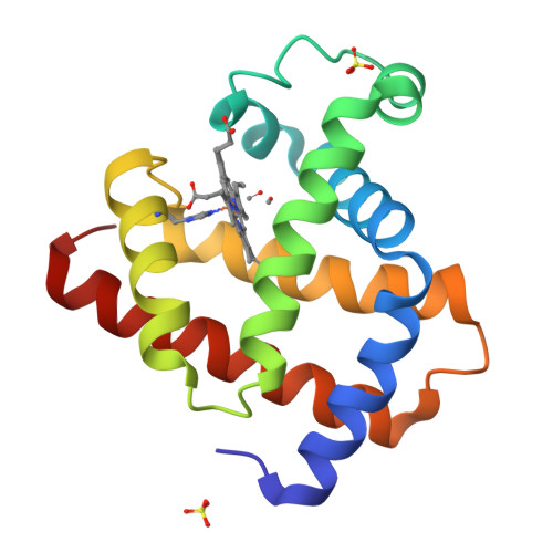
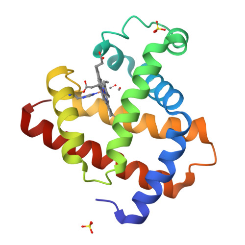
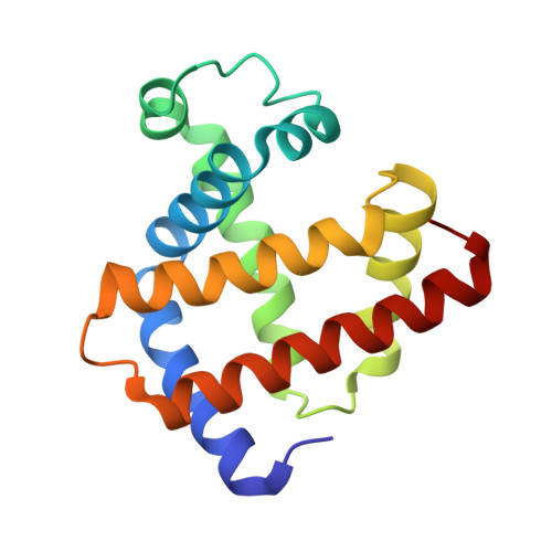
Influence of pump laser fluence on ultrafast myoglobin structural dynamics.
Barends, T.R.M., Gorel, A., Bhattacharyya, S., Schiro, G., Bacellar, C., Cirelli, C., Colletier, J.P., Foucar, L., Grunbein, M.L., Hartmann, E., Hilpert, M., Holton, J.M., Johnson, P.J.M., Kloos, M., Knopp, G., Marekha, B., Nass, K., Nass Kovacs, G., Ozerov, D., Stricker, M., Weik, M., Doak, R.B., Shoeman, R.L., Milne, C.J., Huix-Rotllant, M., Cammarata, M., Schlichting, I.(2024) Nature 626: 905-911
- PubMed: 38355794
- DOI: https://doi.org/10.1038/s41586-024-07032-9
- Primary Citation of Related Structures:
8BKH, 8BKN, 8R8F, 8R8G, 8R8H, 8R8I, 8R8J, 8R8W, 8R8X, 8R8Y, 8R8Z, 8R90, 8R91, 8R92, 8R93, 8R94, 8R95, 8R9C, 8R9D, 8R9E, 8R9F, 8R9G, 8R9H, 8R9I, 8R9J, 8R9K, 8R9L, 8R9M, 8R9N, 8R9P, 8R9Q, 8RA1, 8RA2, 8RA3, 8RA4, 8RA5, 8RA6, 8RA7, 8RA8, 8RA9, 8RAA, 8RAB, 8RAC, 8RAD, 8RAE - PubMed Abstract:
High-intensity femtosecond pulses from an X-ray free-electron laser enable pump-probe experiments for the investigation of electronic and nuclear changes during light-induced reactions. On timescales ranging from femtoseconds to milliseconds and for a variety of biological systems, time-resolved serial femtosecond crystallography (TR-SFX) has provided detailed structural data for light-induced isomerization, breakage or formation of chemical bonds and electron transfer 1,2 . However, all ultrafast TR-SFX studies to date have employed such high pump laser energies that nominally several photons were absorbed per chromophore 3-17 . As multiphoton absorption may force the protein response into non-physiological pathways, it is of great concern 18,19 whether this experimental approach 20 allows valid conclusions to be drawn vis-à-vis biologically relevant single-photon-induced reactions 18,19 . Here we describe ultrafast pump-probe SFX experiments on the photodissociation of carboxymyoglobin, showing that different pump laser fluences yield markedly different results. In particular, the dynamics of structural changes and observed indicators of the mechanistically important coherent oscillations of the Fe-CO bond distance (predicted by recent quantum wavepacket dynamics 21 ) are seen to depend strongly on pump laser energy, in line with quantum chemical analysis. Our results confirm both the feasibility and necessity of performing ultrafast TR-SFX pump-probe experiments in the linear photoexcitation regime. We consider this to be a starting point for reassessing both the design and the interpretation of ultrafast TR-SFX pump-probe experiments 20 such that mechanistically relevant insight emerges.
Organizational Affiliation:
Max Planck Institute for Medical Research, Heidelberg, Germany. Thomas.Barends@mpimf-heidelberg.mpg.de.


