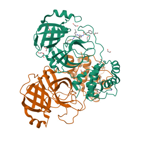
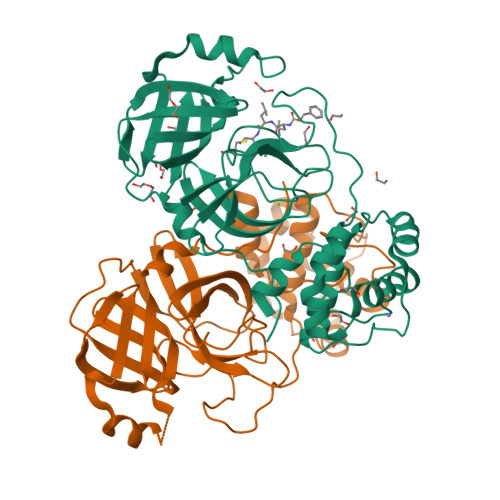
RCSB PDB - 8P86: Crystal structure of the main protease (3CLpro/Mpro) of SARS-CoV-2 obtained in presence of 5 mM MG-132, from an "old" crystal.

 8P86

Crystal structure of the main protease (3CLpro/Mpro) of SARS-CoV-2 obtained in presence of 5 mM MG-132, from an "old" crystal.


Experimental Data Snapshot

  • Method: X-RAY DIFFRACTION
  • Resolution: 1.85 Å
  • R-Value Free: 0.197 
  • R-Value Work: 0.173 
  • R-Value Observed: 0.174 

Starting Model: experimental
View more details

wwPDB Validation   3D Report Full Report


Ligand Structure Quality Assessment 

Created with Raphaël 2.3.0Worse 01 BetterLigand structure goodness of fit to experimental dataBest fitted ALDClick on this verticalbar to view details

This is version 1.1 of the entry. See complete history


Literature

Unexpected Single-Ligand Occupancy and Negative Cooperativity in the SARS-CoV-2 Main Protease.

Albani, S.Costanzi, E.Hoang, G.L.Kuzikov, M.Frings, M.Ansari, N.Demitri, N.Nguyen, T.T.Rizzi, V.Schulz, J.B.Bolm, C.Zaliani, A.Carloni, P.Storici, P.Rossetti, G.

(2024) J Chem Inf Model 64: 892-904

  • DOI: https://doi.org/10.1021/acs.jcim.3c01497
  • Primary Citation of Related Structures:  
    7PHZ, 8P54, 8P55, 8P56, 8P57, 8P58, 8P5A, 8P5B, 8P5C, 8P86, 8P87

  • PubMed Abstract: 

    Many homodimeric enzymes tune their functions by exploiting either negative or positive cooperativity between subunits. In the SARS-CoV-2 Main protease (Mpro) homodimer, the latter has been suggested by symmetry in most of the 500 reported protease/ligand complex structures solved by macromolecular crystallography (MX). Here we apply the latter to both covalent and noncovalent ligands in complex with Mpro. Strikingly, our experiments show that the occupation of both active sites of the dimer originates from an excess of ligands. Indeed, cocrystals obtained using a 1:1 ligand/protomer stoichiometry lead to single occupation only. The empty binding site exhibits a catalytically inactive geometry in solution, as suggested by molecular dynamics simulations. Thus, Mpro operates through negative cooperativity with the asymmetric activity of the catalytic sites. This allows it to function with a wide range of substrate concentrations, making it resistant to saturation and potentially difficult to shut down, all properties advantageous for the virus' adaptability and resistance.


  • Organizational Affiliation

    Institute for Neuroscience and Medicine (INM-9), Forschungszentrum Jülich, Jülich 52425, Germany.


Macromolecules
Find similar proteins by:  (by identity cutoff)  |  3D Structure
Entity ID: 1
MoleculeChains Sequence LengthOrganismDetailsImage
3C-like proteinase nsp5
A, B
306Severe acute respiratory syndrome coronavirus 2Mutation(s): 0 
Gene Names: rep1a-1b
EC: 3.4.22.69
UniProt
Find proteins for P0DTD1 (Severe acute respiratory syndrome coronavirus 2)
Explore P0DTD1 
Go to UniProtKB:  P0DTD1
Entity Groups  
Sequence Clusters30% Identity50% Identity70% Identity90% Identity95% Identity100% Identity
UniProt GroupP0DTD1
Sequence Annotations
Expand
  • Reference Sequence
Small Molecules
Ligands 5 Unique
IDChains Name / Formula / InChI Key2D Diagram3D Interactions
ALD (Subject of Investigation/LOI)
Query on ALD

Download Ideal Coordinates CCD File 
C [auth A]N-[(benzyloxy)carbonyl]-L-leucyl-N-[(2S)-1-hydroxy-4-methylpentan-2-yl]-L-leucinamide
C26 H43 N3 O5
WUJQMWDTZKIKQZ-VABKMULXSA-N
PEG
Query on PEG

Download Ideal Coordinates CCD File 
H [auth A],
M [auth A],
N [auth A]
DI(HYDROXYETHYL)ETHER
C4 H10 O3
MTHSVFCYNBDYFN-UHFFFAOYSA-N
EDO
Query on EDO

Download Ideal Coordinates CCD File 
D [auth A]
E [auth A]
F [auth A]
G [auth A]
I [auth A]
1,2-ETHANEDIOL
C2 H6 O2
LYCAIKOWRPUZTN-UHFFFAOYSA-N
CL
Query on CL

Download Ideal Coordinates CCD File 
Q [auth A]CHLORIDE ION
Cl
VEXZGXHMUGYJMC-UHFFFAOYSA-M
NA
Query on NA

Download Ideal Coordinates CCD File 
O [auth A],
T [auth B]
SODIUM ION
Na
FKNQFGJONOIPTF-UHFFFAOYSA-N
Biologically Interesting Molecules (External Reference) 1 Unique
Experimental Data & Validation

Experimental Data

  • Method: X-RAY DIFFRACTION
  • Resolution: 1.85 Å
  • R-Value Free: 0.197 
  • R-Value Work: 0.173 
  • R-Value Observed: 0.174 
  • Space Group: P 21 21 21
Unit Cell:
Length ( Å )Angle ( ˚ )
a = 68.366α = 90
b = 99.351β = 90
c = 102.801γ = 90
Software Package:
Software NamePurpose
PHENIXrefinement
PHENIXrefinement
XDSdata reduction
Aimlessdata scaling
PHASERphasing

Structure Validation

View Full Validation Report


Ligand Structure Quality Assessment 

Created with Raphaël 2.3.0Worse 01 BetterLigand structure goodness of fit to experimental dataBest fitted ALDClick on this verticalbar to view details

Entry History & Funding Information

Deposition Data


Funding OrganizationLocationGrant Number
European CommissionEuropean Union101003551

Revision History  (Full details and data files)

  • Version 1.0: 2024-05-01
    Type: Initial release
  • Version 1.1: 2024-10-23
    Changes: Structure summary