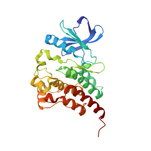
8FD9

Structure of BTK kinase domain with the second-generation inhibitor acalabrutinib


Experimental Data Snapshot

  • Method: X-RAY DIFFRACTION
  • Resolution: 1.70 Å
  • R-Value Free: 0.228 
  • R-Value Work: 0.187 
  • R-Value Observed: 0.189 

Starting Model: experimental
View more details

wwPDB Validation   3D Report Full Report


Ligand Structure Quality Assessment 


This is version 2.2 of the entry. See complete history


Literature

Structure of BTK kinase domain with the second-generation inhibitors acalabrutinib and tirabrutinib.

Lin, D.Y.Andreotti, A.H.

(2023) PLoS One 18: e0290872-e0290872

  • DOI: https://doi.org/10.1371/journal.pone.0290872
  • Primary Citation of Related Structures:  
    8FD9, 8FF0

  • PubMed Abstract: 

    Bruton's tyrosine kinase (BTK) is the target of the therapeutic agent, Ibrutinib, that treats chronic lymphocyte leukemia (CLL), mantle cell lymphoma (MCL) and other B cell malignancies. Ibrutinib is a first in class, covalent BTK inhibitor that limits B-cell survival and proliferation. Designing new inhibitors of BTK has been an important objective for advancing development of improved therapeutic agents against cancer and autoimmune disorders. Based on the success of Ibrutinib, several second-generation irreversible BTK inhibitors have been developed that exhibit fewer off-target effects. However, the binding-mode and their interaction with Btk have not been experimentally determined and evaluated at atomic resolution. Here we determined the first crystal structure of the BTK kinase domain in complex with acalabrutinib. In addition, we report a structure of the BTK/tirabrutinib complex and compare these structures with previously solved structures. The structures provide insight in the superior selectivity reported for acalabrutinb and guide future BTK inhibitor development.


  • Organizational Affiliation

    Roy J. Carver Department of Biochemistry, Biophysics and Molecular Biology, Iowa State, University, Ames, IA, United States of America.


Macromolecules
Find similar proteins by:  (by identity cutoff)  |  3D Structure
Entity ID: 1
MoleculeChains Sequence LengthOrganismDetailsImage
Tyrosine-protein kinase BTK271Mus musculusMutation(s): 8 
Gene Names: BtkBpk
EC: 2.7.10.2
UniProt & NIH Common Fund Data Resources
Find proteins for P35991 (Mus musculus)
Explore P35991 
Go to UniProtKB:  P35991
IMPC:  MGI:88216
Entity Groups  
Sequence Clusters30% Identity50% Identity70% Identity90% Identity95% Identity100% Identity
UniProt GroupP35991
Sequence Annotations
Expand
  • Reference Sequence
Small Molecules
Binding Affinity Annotations 
IDSourceBinding Affinity
XQQ BindingDB:  8FD9 Kd: min: 2.6, max: 21 (nM) from 2 assay(s)
IC50: min: 3, max: 1000 (nM) from 18 assay(s)
EC50: min: 9.2, max: 10 (nM) from 2 assay(s)
Experimental Data & Validation

Experimental Data

  • Method: X-RAY DIFFRACTION
  • Resolution: 1.70 Å
  • R-Value Free: 0.228 
  • R-Value Work: 0.187 
  • R-Value Observed: 0.189 
  • Space Group: P 21 21 21
Unit Cell:
Length ( Å )Angle ( ˚ )
a = 42.921α = 90
b = 50.31β = 90
c = 123.483γ = 90
Software Package:
Software NamePurpose
PHENIXrefinement
autoPROCdata processing
PHASERphasing
autoPROCdata reduction
XDSdata scaling
Cootmodel building

Structure Validation

View Full Validation Report


Ligand Structure Quality Assessment 


Entry History & Funding Information

Deposition Data


Funding OrganizationLocationGrant Number
National Institutes of Health/National Institute Of Allergy and Infectious Diseases (NIH/NIAID)United States--

Revision History  (Full details and data files)

  • Version 1.0: 2023-07-05
    Type: Initial release
  • Version 2.0: 2023-08-23
    Changes: Advisory, Data collection, Derived calculations, Non-polymer description, Structure summary
  • Version 2.1: 2023-09-13
    Changes: Database references
  • Version 2.2: 2024-11-20
    Changes: Structure summary