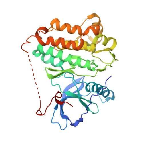
8F1H

EGFR kinase in complex with TAS6417 (CLN-081)


Experimental Data Snapshot

  • Method: X-RAY DIFFRACTION
  • Resolution: 2.80 Å
  • R-Value Free: 0.212 
  • R-Value Work: 0.184 
  • R-Value Observed: 0.186 

wwPDB Validation   3D Report Full Report


Ligand Structure Quality Assessment 


This is version 1.2 of the entry. See complete history


Literature

Biochemical analysis of EGFR exon20 insertion variants insASV and insSVD and their inhibitor sensitivity.

Zhao, H.Beyett, T.S.Jiang, J.Rana, J.K.Schaeffner, I.K.Santana, J.Janne, P.A.Eck, M.J.

(2024) Proc Natl Acad Sci U S A 121: e2417144121-e2417144121

  • DOI: https://doi.org/10.1073/pnas.2417144121
  • Primary Citation of Related Structures:  
    8F1H, 8F1W, 8F1X, 8F1Y, 8F1Z

  • PubMed Abstract: 

    Somatic mutations in the epidermal growth factor receptor (EGFR) are a major cause of non-small cell lung cancer. Among these structurally diverse alterations, exon 20 insertions represent a unique subset that rarely respond to EGFR tyrosine kinase inhibitors (TKIs). Therefore, there is a significant need to develop inhibitors that are active against this class of activating mutations. Here, we conducted biochemical analysis of the two most frequent exon 20 insertion variants, V769_D770insASV (insASV) and D770_N771insSVD (insSVD) to better understand their drug sensitivity and resistance. From kinetic studies, we found that EGFR insASV and insSVD are similarly active, but have lower K m, ATP values compared to the L858R variant, which contributes to their lack of sensitivity to 1st-3rd generation EGFR TKIs. Biochemical, structural, and cellular studies of a diverse panel of EGFR inhibitors revealed that the more recently developed compounds BAY-568, TAS6417, and TAK-788 inhibit EGFR insASV and insSVD in a mutant-selective manner, with BAY-568 being the most potent and selective versus wild-type (WT) EGFR. Cocrystal structures with WT EGFR reveal the binding modes of each of these inhibitors and of poziotinib, a potent but not mutantselective inhibitor, and together they define interactions shared by the mutant-selective agents. Collectively, our results show that these exon20 insertion variants are not inherently inhibitor resistant, rather they differ in their drug sensitivity from WT EGFR. However, they are similar to each other, indicating that a single inhibitor should be effective for several of the diverse exon 20 insertion variants.


  • Organizational Affiliation

    Department of Cancer Biology, Dana-Farber Cancer Institute, Boston, MA 02215.


Macromolecules
Find similar proteins by:  (by identity cutoff)  |  3D Structure
Entity ID: 1
MoleculeChains Sequence LengthOrganismDetailsImage
Epidermal growth factor receptor331Homo sapiensMutation(s): 0 
Gene Names: EGFRERBBERBB1HER1
EC: 2.7.10.1
UniProt & NIH Common Fund Data Resources
Find proteins for P00533 (Homo sapiens)
Explore P00533 
Go to UniProtKB:  P00533
PHAROS:  P00533
GTEx:  ENSG00000146648 
Entity Groups  
Sequence Clusters30% Identity50% Identity70% Identity90% Identity95% Identity100% Identity
UniProt GroupP00533
Sequence Annotations
Expand
  • Reference Sequence
Small Molecules
Ligands 2 Unique
IDChains Name / Formula / InChI Key2D Diagram3D Interactions
X9H (Subject of Investigation/LOI)
Query on X9H

Download Ideal Coordinates CCD File 
B [auth A]N-[(5P,8S,10R)-4-amino-6-methyl-5-(quinolin-3-yl)-8,9-dihydropyrimido[5,4-b]indolizin-8-yl]prop-2-enamide
C23 H20 N6 O
MKCYPWYURWOKST-INIZCTEOSA-N
CIT
Query on CIT

Download Ideal Coordinates CCD File 
C [auth A]CITRIC ACID
C6 H8 O7
KRKNYBCHXYNGOX-UHFFFAOYSA-N
Binding Affinity Annotations 
IDSourceBinding Affinity
X9H BindingDB:  8F1H IC50: 1.1 (nM) from 1 assay(s)
Experimental Data & Validation

Experimental Data

  • Method: X-RAY DIFFRACTION
  • Resolution: 2.80 Å
  • R-Value Free: 0.212 
  • R-Value Work: 0.184 
  • R-Value Observed: 0.186 
  • Space Group: I 2 3
Unit Cell:
Length ( Å )Angle ( ˚ )
a = 146.317α = 90
b = 146.317β = 90
c = 146.317γ = 90
Software Package:
Software NamePurpose
xia2data scaling
PHENIXrefinement
xia2data reduction
PHASERphasing
Cootmodel building

Structure Validation

View Full Validation Report


Ligand Structure Quality Assessment 


Entry History & Funding Information

Deposition Data


Funding OrganizationLocationGrant Number
National Institutes of Health/National Cancer Institute (NIH/NCI)United States5R01CA116020-16

Revision History  (Full details and data files)

  • Version 1.0: 2023-11-08
    Type: Initial release
  • Version 1.1: 2024-10-23
    Changes: Structure summary
  • Version 1.2: 2024-11-06
    Changes: Database references