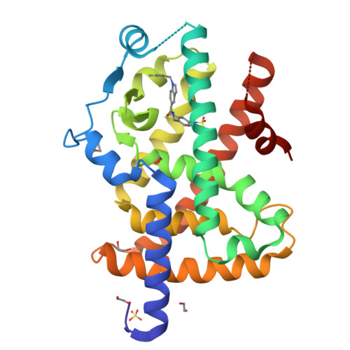
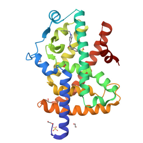
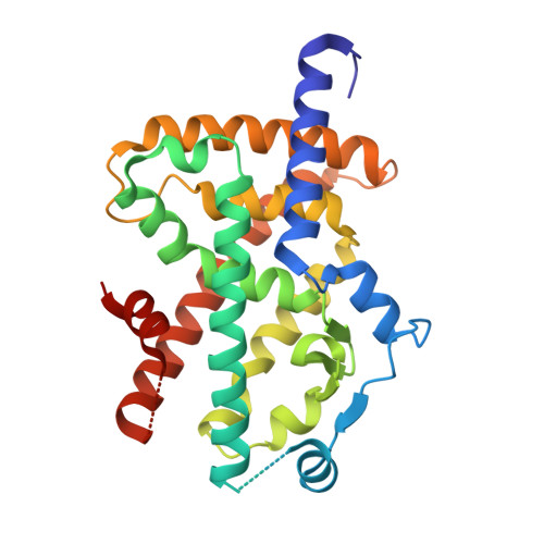
RCSB PDB - 7P4E: Crystal structure of PPARgamma in complex with compound FL217

 7P4E

Crystal structure of PPARgamma in complex with compound FL217


Experimental Data Snapshot

  • Method: X-RAY DIFFRACTION
  • Resolution: 2.40 Å
  • R-Value Free: 0.268 
  • R-Value Work: 0.229 
  • R-Value Observed: 0.231 

Starting Model: experimental
View more details

wwPDB Validation   3D Report Full Report


Ligand Structure Quality Assessment 

Created with Raphaël 2.3.0Worse 01 BetterLigand structure goodness of fit to experimental dataBest fitted 5IVClick on this verticalbar to view details

This is version 1.1 of the entry. See complete history


Literature

Structure-Based Design of Dual Partial Peroxisome Proliferator-Activated Receptor gamma Agonists/Soluble Epoxide Hydrolase Inhibitors.

Lillich, F.F.Willems, S.Ni, X.Kilu, W.Borkowsky, C.Brodsky, M.Kramer, J.S.Brunst, S.Hernandez-Olmos, V.Heering, J.Schierle, S.Kestner, R.I.Mayser, F.M.Helmstadter, M.Gobel, T.Weizel, L.Namgaladze, D.Kaiser, A.Steinhilber, D.Pfeilschifter, W.Kahnt, A.S.Proschak, A.Chaikuad, A.Knapp, S.Merk, D.Proschak, E.

(2021) J Med Chem 64: 17259-17276

  • DOI: https://doi.org/10.1021/acs.jmedchem.1c01331
  • Primary Citation of Related Structures:  
    7P4E, 7P4K

  • PubMed Abstract: 

    Polypharmaceutical regimens often impair treatment of patients with metabolic syndrome (MetS), a complex disease cluster, including obesity, hypertension, heart disease, and type II diabetes. Simultaneous targeting of soluble epoxide hydrolase (sEH) and peroxisome proliferator-activated receptor γ (PPARγ) synergistically counteracted MetS in various in vivo models, and dual sEH inhibitors/PPARγ agonists hold great potential to reduce the problems associated with polypharmacy in the context of MetS. However, full activation of PPARγ leads to fluid retention associated with edema and weight gain, while partial PPARγ agonists do not have these drawbacks. In this study, we designed a dual partial PPARγ agonist/sEH inhibitor using a structure-guided approach. Exhaustive structure-activity relationship studies lead to the successful optimization of the designed lead. Crystal structures of one representative compound with both targets revealed potential points for optimization. The optimized compounds exhibited favorable metabolic stability, toxicity, selectivity, and desirable activity in adipocytes and macrophages.


  • Organizational Affiliation

    Institute of Pharmaceutical Chemistry, Goethe-University, Max-von-Laue-Str. 9, D-60438 Frankfurt am Main, Germany.


Macromolecules
Find similar proteins by:  (by identity cutoff)  |  3D Structure
Entity ID: 1
MoleculeChains Sequence LengthOrganismDetailsImage
Peroxisome proliferator-activated receptor gamma277Homo sapiensMutation(s): 0 
Gene Names: PPARGNR1C3
UniProt & NIH Common Fund Data Resources
Find proteins for P37231 (Homo sapiens)
Explore P37231 
Go to UniProtKB:  P37231
PHAROS:  P37231
GTEx:  ENSG00000132170 
Entity Groups  
Sequence Clusters30% Identity50% Identity70% Identity90% Identity95% Identity100% Identity
UniProt GroupP37231
Sequence Annotations
Expand
  • Reference Sequence
Small Molecules
Ligands 3 Unique
IDChains Name / Formula / InChI Key2D Diagram3D Interactions
5IV (Subject of Investigation/LOI)
Query on 5IV

Download Ideal Coordinates CCD File 
B [auth A]~{N}-[[4-(cyclopropylsulfonylamino)-2-(trifluoromethyl)phenyl]methyl]-1-[(3-fluorophenyl)methyl]indole-5-carboxamide
C27 H23 F4 N3 O3 S
NONHTXRHOREFAH-UHFFFAOYSA-N
SO4
Query on SO4

Download Ideal Coordinates CCD File 
C [auth A]SULFATE ION
O4 S
QAOWNCQODCNURD-UHFFFAOYSA-L
EDO
Query on EDO

Download Ideal Coordinates CCD File 
D [auth A]
E [auth A]
F [auth A]
G [auth A]
H [auth A]
1,2-ETHANEDIOL
C2 H6 O2
LYCAIKOWRPUZTN-UHFFFAOYSA-N
Binding Affinity Annotations 
IDSourceBinding Affinity
5IV BindingDB:  7P4E EC50: min: 180, max: 220 (nM) from 2 assay(s)
Experimental Data & Validation

Experimental Data

  • Method: X-RAY DIFFRACTION
  • Resolution: 2.40 Å
  • R-Value Free: 0.268 
  • R-Value Work: 0.229 
  • R-Value Observed: 0.231 
  • Space Group: P 41 21 2
Unit Cell:
Length ( Å )Angle ( ˚ )
a = 63.013α = 90
b = 63.013β = 90
c = 167.621γ = 90
Software Package:
Software NamePurpose
Aimlessdata scaling
REFMACrefinement
PDB_EXTRACTdata extraction
XDSdata reduction
PHASERphasing

Structure Validation

View Full Validation Report


Ligand Structure Quality Assessment 

Created with Raphaël 2.3.0Worse 01 BetterLigand structure goodness of fit to experimental dataBest fitted 5IVClick on this verticalbar to view details

Entry History 

Deposition Data

Revision History  (Full details and data files)

  • Version 1.0: 2022-07-06
    Type: Initial release
  • Version 1.1: 2024-01-31
    Changes: Data collection, Refinement description