6HK5

X-ray structure of a truncated mutant of the metallochaperone CooJ with a high-affinity nickel-binding site


Experimental Data Snapshot

  • Method: X-RAY DIFFRACTION
  • Resolution: 2.04 Å
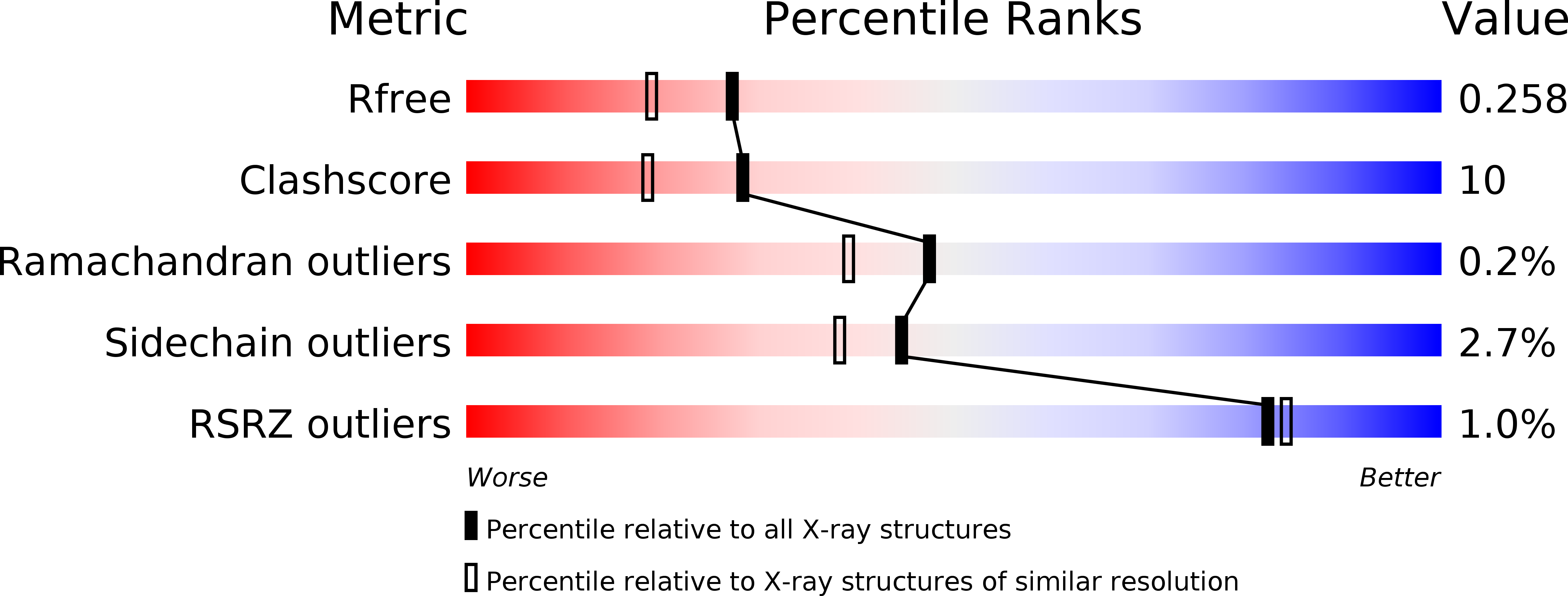
  • R-Value Free: 0.259 
  • R-Value Work: 0.200 
  • R-Value Observed: 0.202 

wwPDB Validation   3D Report Full Report


Ligand Structure Quality Assessment 


This is version 1.2 of the entry. See complete history


Literature

The carbon monoxide dehydrogenase accessory protein CooJ is a histidine-rich multidomain dimer containing an unexpected Ni(II)-binding site.

Alfano, M.Perard, J.Carpentier, P.Basset, C.Zambelli, B.Timm, J.Crouzy, S.Ciurli, S.Cavazza, C.

(2019) J Biol Chem 294: 7601-7614

  • DOI: https://doi.org/10.1074/jbc.RA119.008011
  • Primary Citation of Related Structures:  
    6HK5

  • PubMed Abstract: 

    Activation of nickel enzymes requires specific accessory proteins organized in multiprotein complexes controlling metal transfer to the active site. Histidine-rich clusters are generally present in at least one of the metallochaperones involved in nickel delivery. The maturation of carbon monoxide dehydrogenase in the proteobacterium Rhodospirillum rubrum requires three accessory proteins, CooC, CooT, and CooJ, dedicated to nickel insertion into the active site, a distorted [NiFe 3 S 4 ] cluster coordinated to an iron site. Previously, CooJ from R. rubrum ( Rr CooJ) has been described as a nickel chaperone with 16 histidines and 2 cysteines at its C terminus. Here, the X-ray structure of a truncated version of Rr CooJ, combined with small-angle X-ray scattering data and a modeling study of the full-length protein, revealed a homodimer comprising a coiled coil with two independent and highly flexible His tails. Using isothermal calorimetry, we characterized several metal-binding sites (four per dimer) involving the His-rich motifs and having similar metal affinity ( K D = 1.6 μm). Remarkably, biophysical approaches, site-directed mutagenesis, and X-ray crystallography uncovered an additional nickel-binding site at the dimer interface, which binds Ni(II) with an affinity of 380 nm Although Rr CooJ was initially thought to be a unique protein, a proteome database search identified at least 46 bacterial CooJ homologs. These homologs all possess two spatially separated nickel-binding motifs: a variable C-terminal histidine tail and a strictly conserved H(W/F) X 2 H X 3 H motif, identified in this study, suggesting a dual function for CooJ both as a nickel chaperone and as a nickel storage protein.


  • Organizational Affiliation

    From the Laboratory of Chemistry and Biology of Metals, Université Grenoble Alpes, CEA, CNRS, F-38000 Grenoble, France and.


Macromolecules
Find similar proteins by:  (by identity cutoff)  |  3D Structure
Entity ID: 1
MoleculeChains Sequence LengthOrganismDetailsImage
CooJ68Rhodospirillum rubrumMutation(s): 0 
Gene Names: cooJ
UniProt
Find proteins for P72321 (Rhodospirillum rubrum)
Explore P72321 
Go to UniProtKB:  P72321
Entity Groups  
Sequence Clusters30% Identity50% Identity70% Identity90% Identity95% Identity100% Identity
UniProt GroupP72321
Sequence Annotations
Expand
  • Reference Sequence
Small Molecules
Ligands 6 Unique
IDChains Name / Formula / InChI Key2D Diagram3D Interactions
Z3P
Query on Z3P

Download Ideal Coordinates CCD File 
U [auth D]3,3',3''-phosphoryltripropanoic acid
C9 H15 O7 P
XJGZCDJZBUBTKW-UHFFFAOYSA-N
PGE
Query on PGE

Download Ideal Coordinates CCD File 
Q [auth A]TRIETHYLENE GLYCOL
C6 H14 O4
ZIBGPFATKBEMQZ-UHFFFAOYSA-N
PEG
Query on PEG

Download Ideal Coordinates CCD File 
J [auth B],
R [auth A],
S [auth A],
T [auth D]
DI(HYDROXYETHYL)ETHER
C4 H10 O3
MTHSVFCYNBDYFN-UHFFFAOYSA-N
NI
Query on NI

Download Ideal Coordinates CCD File 
I [auth B],
O [auth C],
P [auth A],
V [auth D]
NICKEL (II) ION
Ni
VEQPNABPJHWNSG-UHFFFAOYSA-N
CA
Query on CA

Download Ideal Coordinates CCD File 
L [auth C]CALCIUM ION
Ca
BHPQYMZQTOCNFJ-UHFFFAOYSA-N
CL
Query on CL

Download Ideal Coordinates CCD File 
K [auth B],
M [auth C],
N [auth C]
CHLORIDE ION
Cl
VEXZGXHMUGYJMC-UHFFFAOYSA-M
Modified Residues  1 Unique
IDChains TypeFormula2D DiagramParent
MSE
Query on MSE
A [auth B]
B [auth C]
C [auth E]
D [auth G]
E [auth A]
A [auth B],
B [auth C],
C [auth E],
D [auth G],
E [auth A],
F [auth D],
G [auth F],
H
L-PEPTIDE LINKINGC5 H11 N O2 SeMET
Experimental Data & Validation

Experimental Data

  • Method: X-RAY DIFFRACTION
  • Resolution: 2.04 Å
  • R-Value Free: 0.259 
  • R-Value Work: 0.200 
  • R-Value Observed: 0.202 
  • Space Group: P 1 21 1
Unit Cell:
Length ( Å )Angle ( ˚ )
a = 46.8α = 90
b = 74.28β = 92.74
c = 70.64γ = 90
Software Package:
Software NamePurpose
PHENIXrefinement
XDSdata reduction
XSCALEdata scaling
SHELXDphasing

Structure Validation

View Full Validation Report


Ligand Structure Quality Assessment 


Entry History 

Deposition Data

Revision History  (Full details and data files)

  • Version 1.0: 2019-03-27
    Type: Initial release
  • Version 1.1: 2019-10-16
    Changes: Data collection, Database references
  • Version 1.2: 2024-11-13
    Changes: Advisory, Data collection, Database references, Derived calculations, Structure summary