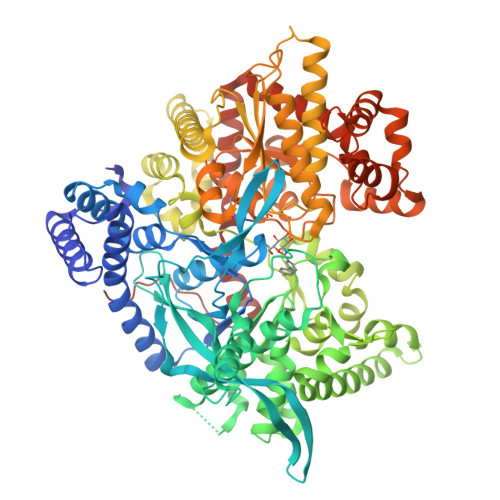
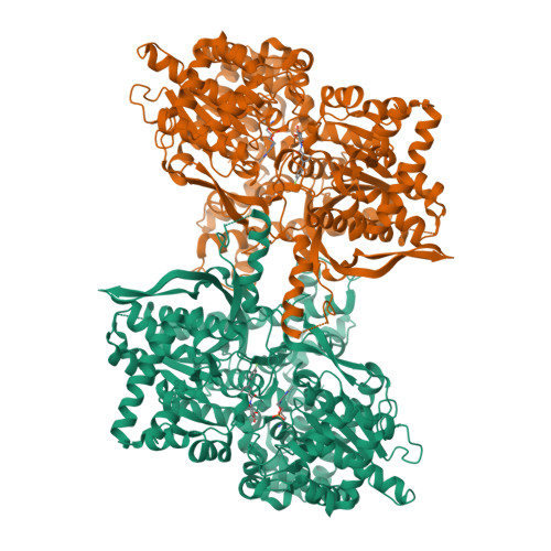
6R0H | pdb_00006r0h

Glycogen Phosphorylase b in complex with 3


Experimental Data Snapshot

  • Method: X-RAY DIFFRACTION
  • Resolution: 2.50 Å
  • R-Value Free: 
    0.213 (Depositor), 0.220 (DCC) 
  • R-Value Work: 
    0.159 (Depositor), 0.170 (DCC) 
  • R-Value Observed: 
    0.162 (Depositor) 

wwPDB Validation   3D Report Full Report


Ligand Structure Quality Assessment 

Created with Raphaël 2.3.0Worse 01 BetterLigand structure goodness of fit to experimental dataBest fitted JN2Click on this verticalbar to view detailsBest fitted PLPClick on this verticalbar to view details

This is version 1.1 of the entry. See complete history


Literature

High Consistency of Structure-Based Design and X-Ray Crystallography: Design, Synthesis, Kinetic Evaluation and Crystallographic Binding Mode Determination of Biphenyl-N-acyl-beta-d-Glucopyranosylamines as Glycogen Phosphorylase Inhibitors.

Fischer, T.Koulas, S.M.Tsagkarakou, A.S.Kyriakis, E.Stravodimos, G.A.Skamnaki, V.T.Liggri, P.G.V.Zographos, S.E.Riedl, R.Leonidas, D.D.

(2019) Molecules 24

  • DOI: https://doi.org/10.3390/molecules24071322
  • Primary Citation of Related Structures:  
    6R0H, 6R0I

  • PubMed Abstract: 

    Structure-based design and synthesis of two biphenyl- N -acyl-β-d-glucopyranosylamine derivatives as well as their assessment as inhibitors of human liver glycogen phosphorylase (hlGPa, a pharmaceutical target for type 2 diabetes) is presented. X-ray crystallography revealed the importance of structural water molecules and that the inhibitory efficacy correlates with the degree of disturbance caused by the inhibitor binding to a loop crucial for the catalytic mechanism. The in silico-derived models of the binding mode generated during the design process corresponded very well with the crystallographic data.


  • Organizational Affiliation

    Institute of Chemistry and Biotechnology, Center of Organic and Medicinal Chemistry, Zurich University of Applied Sciences, Einsiedlerstrasse 31, 8820 Wädenswil, Switzerland. thomas.fischer@zhaw.ch.


Macromolecules
Find similar proteins by:  (by identity cutoff)  |  3D Structure
Entity ID: 1
MoleculeChains Sequence LengthOrganismDetailsImage
Glycogen phosphorylase, muscle form843Oryctolagus cuniculusMutation(s): 0 
EC: 2.4.1.1
UniProt
Find proteins for P00489 (Oryctolagus cuniculus)
Explore P00489 
Go to UniProtKB:  P00489
Entity Groups  
Sequence Clusters30% Identity50% Identity70% Identity90% Identity95% Identity100% Identity
UniProt GroupP00489
Sequence Annotations
Expand
  • Reference Sequence
Small Molecules
Ligands 2 Unique
IDChains Name / Formula / InChI Key2D Diagram3D Interactions
JN2
Query on JN2

Download Ideal Coordinates CCD File 
B [auth A]3-(4-fluorophenyl)-~{N}-[(2~{R},3~{R},4~{S},5~{S},6~{R})-6-(hydroxymethyl)-3,4,5-tris(oxidanyl)oxan-2-yl]benzamide
C19 H20 F N O6
ZNFXGMNTWYBCJQ-OGJJZOIMSA-N
PLP
Query on PLP

Download Ideal Coordinates CCD File 
C [auth A]PYRIDOXAL-5'-PHOSPHATE
C8 H10 N O6 P
NGVDGCNFYWLIFO-UHFFFAOYSA-N
Experimental Data & Validation

Experimental Data

  • Method: X-RAY DIFFRACTION
  • Resolution: 2.50 Å
  • R-Value Free:  0.213 (Depositor), 0.220 (DCC) 
  • R-Value Work:  0.159 (Depositor), 0.170 (DCC) 
  • R-Value Observed: 0.162 (Depositor) 
Space Group: P 43 21 2
Unit Cell:
Length ( Å )Angle ( ˚ )
a = 128.287α = 90
b = 128.287β = 90
c = 116.12γ = 90
Software Package:
Software NamePurpose
REFMACrefinement
CrysalisProdata reduction
Aimlessdata scaling

Structure Validation

View Full Validation Report


Ligand Structure Quality Assessment 

Created with Raphaël 2.3.0Worse 01 BetterLigand structure goodness of fit to experimental dataBest fitted JN2Click on this verticalbar to view detailsBest fitted PLPClick on this verticalbar to view details

Entry History 

Deposition Data

Revision History  (Full details and data files)

  • Version 1.0: 2019-04-10
    Type: Initial release
  • Version 1.1: 2019-04-24
    Changes: Data collection, Database references