6QN0

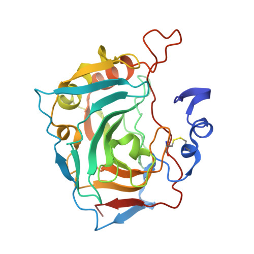
Three dimensional structure of human carbonic anhydrase XII in complex with benzenesulfonamide


Experimental Data Snapshot

  • Method: X-RAY DIFFRACTION
  • Resolution: 1.89 Å
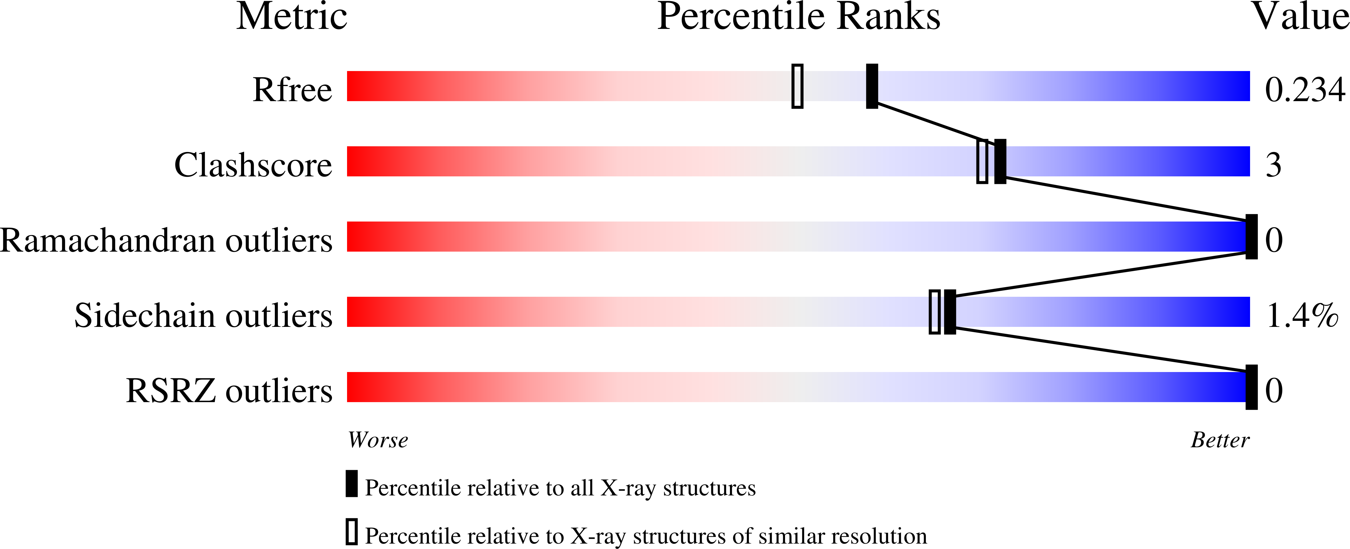
  • R-Value Free: 0.225 
  • R-Value Work: 0.165 
  • R-Value Observed: 0.168 

Starting Model: experimental
View more details

wwPDB Validation   3D Report Full Report


Ligand Structure Quality Assessment 


This is version 1.5 of the entry. See complete history


Literature

Halogenated and di-substituted benzenesulfonamides as selective inhibitors of carbonic anhydrase isoforms.

Zaksauskas, A.Capkauskaite, E.Jezepcikas, L.Linkuviene, V.Paketuryte, V.Smirnov, A.Leitans, J.Kazaks, A.Dvinskis, E.Manakova, E.Grazulis, S.Tars, K.Matulis, D.

(2020) Eur J Med Chem 185: 111825-111825

  • DOI: https://doi.org/10.1016/j.ejmech.2019.111825
  • Primary Citation of Related Structures:  
    6QN0, 6QN2, 6QN5, 6QN6, 6QNL, 6QUT, 6R6F, 6R6J, 6R6Y, 6R71

  • PubMed Abstract: 

    By applying an approach of a "ring with two tails", a series of novel inhibitors possessing high-affinity and significant selectivity towards selected carbonic anhydrase (CA) isoforms has been designed. The "ring" consists of 2-chloro/bromo-benzenesulfonamide, where the sulfonamide group is as an anchor coordinating the Zn(II) in the active site of CAs, and halogen atom orients the ring affecting the affinity and selectivity. First "tail" is a substituent containing carbonyl, carboxyl, hydroxyl, ether groups or hydrophilic amide linkage. The second "tail" contains aryl- or alkyl-substituents attached through a sulfanyl or sulfonyl group. Both "tails" are connected to the benzene ring and play a crucial role in selectivity. Varying the substituents, we designed compounds selective for CA VII, CA IX, CA XII, or CA XIV. Since due to binding-linked protonation reactions the binding-ready fractions of the compound and protein are much lower than one, the "intrinsic" affinities were calculated that should be used to study correlations between crystal structures and the thermodynamics of binding for rational drug design. The "intrinsic" affinities together with the intrinsic enthalpies and entropies of binding together with co-crystal structures were used demonstrate structural factors determining major contributions for compound affinity and selectivity.


  • Organizational Affiliation

    Department of Biothermodynamics and Drug Design, Institute of Biotechnology, Life Sciences Center, Vilnius University, Saulėtekio al. 7, Vilnius, LT, 10257, Lithuania.


Macromolecules
Find similar proteins by:  (by identity cutoff)  |  3D Structure
Entity ID: 1
MoleculeChains Sequence LengthOrganismDetailsImage
Carbonic anhydrase 12
A, B, C, D
263Homo sapiensMutation(s): 0 
Gene Names: CA12
EC: 4.2.1.1
UniProt & NIH Common Fund Data Resources
Find proteins for O43570 (Homo sapiens)
Explore O43570 
Go to UniProtKB:  O43570
PHAROS:  O43570
GTEx:  ENSG00000074410 
Entity Groups  
Sequence Clusters30% Identity50% Identity70% Identity90% Identity95% Identity100% Identity
UniProt GroupO43570
Sequence Annotations
Expand
  • Reference Sequence
Small Molecules
Ligands 2 Unique
IDChains Name / Formula / InChI Key2D Diagram3D Interactions
J8Q (Subject of Investigation/LOI)
Query on J8Q

Download Ideal Coordinates CCD File 
F [auth A],
H [auth B],
J [auth C],
L [auth D]
~{N}-butyl-4-chloranyl-2-(2-phenylethylsulfanyl)-5-sulfamoyl-benzamide
C19 H23 Cl N2 O3 S2
GKEVLRUIWRFAPB-UHFFFAOYSA-N
ZN
Query on ZN

Download Ideal Coordinates CCD File 
E [auth A],
G [auth B],
I [auth C],
K [auth D]
ZINC ION
Zn
PTFCDOFLOPIGGS-UHFFFAOYSA-N
Binding Affinity Annotations 
IDSourceBinding Affinity
J8Q BindingDB:  6QN0 Kd: min: 3.00e-3, max: 1.4 (nM) from 2 assay(s)
Experimental Data & Validation

Experimental Data

  • Method: X-RAY DIFFRACTION
  • Resolution: 1.89 Å
  • R-Value Free: 0.225 
  • R-Value Work: 0.165 
  • R-Value Observed: 0.168 
  • Space Group: P 1
Unit Cell:
Length ( Å )Angle ( ˚ )
a = 45.76α = 90.02
b = 74.76β = 74.32
c = 76.23γ = 90
Software Package:
Software NamePurpose
REFMACrefinement
SCALAdata scaling

Structure Validation

View Full Validation Report


Ligand Structure Quality Assessment 


Entry History 

Deposition Data

Revision History  (Full details and data files)

  • Version 1.0: 2020-02-26
    Type: Initial release
  • Version 1.1: 2020-03-18
    Changes: Derived calculations
  • Version 1.2: 2021-10-06
    Changes: Data collection, Database references, Derived calculations, Structure summary
  • Version 1.3: 2021-10-13
    Changes: Data collection
  • Version 1.4: 2024-01-24
    Changes: Data collection, Refinement description
  • Version 1.5: 2024-11-06
    Changes: Structure summary