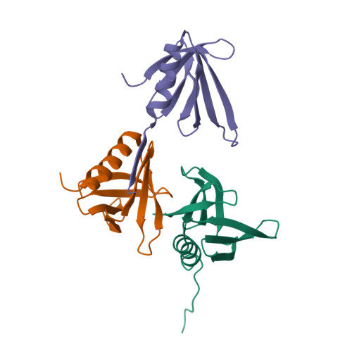
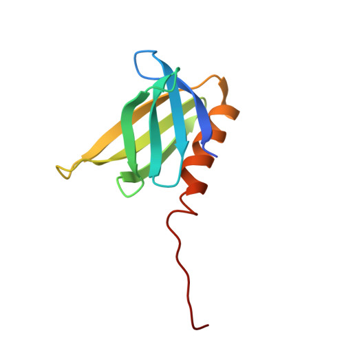
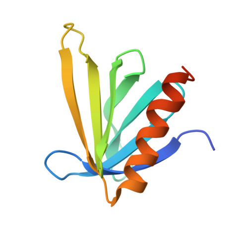
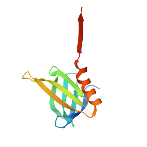
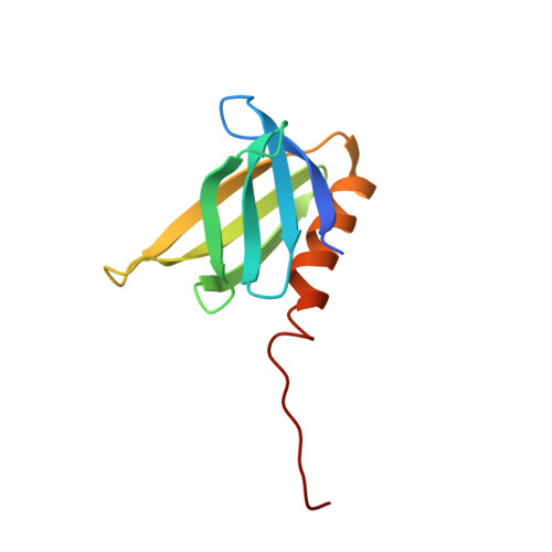
Structural analysis of the LDL receptor-interacting FERM domain in the E3 ubiquitin ligase IDOL reveals an obscured substrate-binding site.
Martinelli, L., Adamopoulos, A., Johansson, P., Wan, P.T., Gunnarsson, J., Guo, H., Boyd, H., Zelcer, N., Sixma, T.K.(2020) J Biological Chem 295: 13570-13583
- PubMed: 32727844
- DOI: https://doi.org/10.1074/jbc.RA120.014349
- Primary Citation of Related Structures:
6QLY, 6QLZ - PubMed Abstract:
Hepatic abundance of the low-density lipoprotein receptor (LDLR) is a critical determinant of circulating plasma LDL cholesterol levels and hence development of coronary artery disease. The sterol-responsive E3 ubiquitin ligase inducible degrader of the LDLR (IDOL) specifically promotes ubiquitination and subsequent lysosomal degradation of the LDLR and thus controls cellular LDL uptake. IDOL contains an extended N-terminal FERM (4.1 protein, ezrin, radixin, and moesin) domain, responsible for substrate recognition and plasma membrane association, and a second C-terminal RING domain, responsible for the E3 ligase activity and homodimerization. As IDOL is a putative lipid-lowering drug target, we investigated the molecular details of its substrate recognition. We produced and isolated full-length IDOL protein, which displayed high autoubiquitination activity. However, in vitro ubiquitination of its substrate, the intracellular tail of the LDLR, was low. To investigate the structural basis for this, we determined crystal structures of the extended FERM domain of IDOL and multiple conformations of its F3ab subdomain. These reveal the archetypal F1-F2-F3 trilobed FERM domain structure but show that the F3c subdomain orientation obscures the target-binding site. To substantiate this finding, we analyzed the full-length FERM domain and a series of truncated FERM constructs by small-angle X-ray scattering (SAXS). The scattering data support a compact and globular core FERM domain with a more flexible and extended C-terminal region. This flexibility may explain the low activity in vitro and suggests that IDOL may require activation for recognition of the LDLR.
Organizational Affiliation:
Division of Biochemistry, Netherlands Cancer Institute, Amsterdam, The Netherlands; Department of Medical Biochemistry, Amsterdam UMC, Amsterdam Cardiovascular Sciences and Gastroenterology and Metabolism, University of Amsterdam, Amsterdam, the Netherlands.


