6EGT | pdb_00006egt

Structure of RVFV envelope protein Gc in postfusion conformation in complex with MES


Experimental Data Snapshot

  • Method: X-RAY DIFFRACTION
  • Resolution: 2.50 Å
  • R-Value Free: 
    0.235 (Depositor), 0.240 (DCC) 
  • R-Value Work: 
    0.196 (Depositor), 0.200 (DCC) 
  • R-Value Observed: 
    0.197 (Depositor) 

Starting Model: experimental
View more details

wwPDB Validation   3D Report Full Report


Ligand Structure Quality Assessment 

Created with Raphaël 2.3.0Worse 01 BetterLigand structure goodness of fit to experimental dataBest fitted NAGClick on this verticalbar to view details

This is version 2.2 of the entry. See complete history


Literature

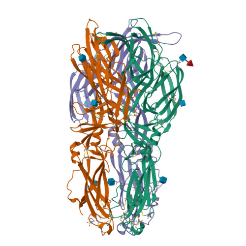
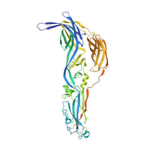
A glycerophospholipid-specific pocket in the RVFV class II fusion protein drives target membrane insertion.

Guardado-Calvo, P.Atkovska, K.Jeffers, S.A.Grau, N.Backovic, M.Perez-Vargas, J.de Boer, S.M.Tortorici, M.A.Pehau-Arnaudet, G.Lepault, J.England, P.Rottier, P.J.Bosch, B.J.Hub, J.S.Rey, F.A.

(2017) Science 358: 663-667

  • DOI: https://doi.org/10.1126/science.aal2712
  • Primary Citation of Related Structures:  
    6EGT, 6EGU

  • PubMed Abstract: 

    The Rift Valley fever virus (RVFV) is transmitted by infected mosquitoes, causing severe disease in humans and livestock across Africa. We determined the x-ray structure of the RVFV class II fusion protein Gc in its postfusion form and in complex with a glycerophospholipid (GPL) bound in a conserved cavity next to the fusion loop. Site-directed mutagenesis and molecular dynamics simulations further revealed a built-in motif allowing en bloc insertion of the fusion loop into membranes, making few nonpolar side-chain interactions with the aliphatic moiety and multiple polar interactions with lipid head groups upon membrane restructuring. The GPL head-group recognition pocket is conserved in the fusion proteins of other arthropod-borne viruses, such as Zika and chikungunya viruses, which have recently caused major epidemics worldwide.


  • Organizational Affiliation

    Institut Pasteur, Département de Virologie, Unité de Virologie Structurale, 75724 Paris Cedex 15, France. pablo.guardado-calvo@pasteur.fr jhub@gwdg.de felix.rey@pasteur.fr.


Macromolecules
Find similar proteins by:  (by identity cutoff)  |  3D Structure
Entity ID: 1
MoleculeChains Sequence LengthOrganismDetailsImage
Glycoprotein
A, B, C
531Rift Valley fever virusMutation(s): 1 
UniProt
Find proteins for P03518 (Rift valley fever virus)
Explore P03518 
Go to UniProtKB:  P03518
Entity Groups  
Sequence Clusters30% Identity50% Identity70% Identity90% Identity95% Identity100% Identity
UniProt GroupP03518
Glycosylation
Glycosylation Sites: 3
Sequence Annotations
Expand
  • Reference Sequence
Oligosaccharides

Help

Entity ID: 2
MoleculeChains Length2D Diagram Glycosylation3D Interactions
beta-L-fucopyranose-(1-6)-2-acetamido-2-deoxy-beta-D-glucopyranose
D
2N-Glycosylation
Glycosylation Resources
GlyTouCan:  G61843VN
GlyCosmos:  G61843VN
GlyGen:  G61843VN
Small Molecules
Ligands 2 Unique
IDChains Name / Formula / InChI Key2D Diagram3D Interactions
NAG
Query on NAG

Download Ideal Coordinates CCD File 
E [auth A]
F [auth A]
G [auth B]
H [auth B]
I [auth B]
2-acetamido-2-deoxy-beta-D-glucopyranose
C8 H15 N O6
OVRNDRQMDRJTHS-FMDGEEDCSA-N
MES
Query on MES

Download Ideal Coordinates CCD File 
J [auth B],
N [auth C]
2-(N-MORPHOLINO)-ETHANESULFONIC ACID
C6 H13 N O4 S
SXGZJKUKBWWHRA-UHFFFAOYSA-N
Experimental Data & Validation

Experimental Data

  • Method: X-RAY DIFFRACTION
  • Resolution: 2.50 Å
  • R-Value Free:  0.235 (Depositor), 0.240 (DCC) 
  • R-Value Work:  0.196 (Depositor), 0.200 (DCC) 
  • R-Value Observed: 0.197 (Depositor) 
Space Group: P 2 21 21
Unit Cell:
Length ( Å )Angle ( ˚ )
a = 72.306α = 90
b = 102.03β = 90
c = 198.892γ = 90
Software Package:
Software NamePurpose
PHENIXrefinement
XDSdata reduction
Aimlessdata scaling
PHASERphasing

Structure Validation

View Full Validation Report


Ligand Structure Quality Assessment 

Created with Raphaël 2.3.0Worse 01 BetterLigand structure goodness of fit to experimental dataBest fitted NAGClick on this verticalbar to view details

Entry History 

Deposition Data

Revision History  (Full details and data files)

  • Version 1.0: 2017-11-01
    Type: Initial release
  • Version 1.1: 2017-11-15
    Changes: Database references
  • Version 1.2: 2019-06-12
    Changes: Data collection, Structure summary
  • Version 2.0: 2020-07-29
    Type: Remediation
    Reason: Carbohydrate remediation
    Changes: Atomic model, Data collection, Derived calculations, Structure summary
  • Version 2.1: 2024-01-17
    Changes: Data collection, Database references, Derived calculations, Refinement description, Structure summary
  • Version 2.2: 2024-11-06
    Changes: Structure summary