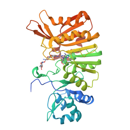
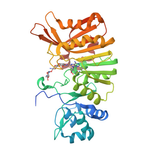
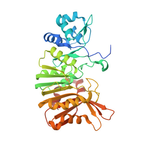
RCSB PDB - 5U1E: Crystal structure of a methyltransferase involved in the biosynthesis of gentamicin in complex with the kanamycin B

 5U1E

Crystal structure of a methyltransferase involved in the biosynthesis of gentamicin in complex with the kanamycin B


Experimental Data Snapshot

  • Method: X-RAY DIFFRACTION
  • Resolution: 2.21 Å
  • R-Value Free: 0.247 
  • R-Value Work: 0.189 
  • R-Value Observed: 0.195 

Starting Model: experimental
View more details

wwPDB Validation   3D Report Full Report


Ligand Structure Quality Assessment 

Created with Raphaël 2.3.0Worse 01 BetterLigand structure goodness of fit to experimental dataBest fitted 9CSClick on this verticalbar to view detailsBest fitted SAHClick on this verticalbar to view details

This is version 1.5 of the entry. See complete history


Literature

Structural Basis of the Selectivity of GenN, an Aminoglycoside N-Methyltransferase Involved in Gentamicin Biosynthesis.

Bury, P.D.S.Huang, F.Li, S.Sun, Y.Leadlay, P.F.Dias, M.V.B.

(2017) ACS Chem Biol 12: 2779-2787

  • DOI: https://doi.org/10.1021/acschembio.7b00466
  • Primary Citation of Related Structures:  
    5TYQ, 5U0N, 5U0T, 5U18, 5U19, 5U1E, 5U1I, 5U4T

  • PubMed Abstract: 

    Gentamicins are heavily methylated, clinically valuable pseudotrisaccharide antibiotics produced by Micromonospora echinospora. GenN has been characterized as an S-adenosyl-l-methionine-dependent methyltransferase with low sequence similarity to other enzymes. It is responsible for the 3″-N-methylation of 3″-dehydro-3″-amino-gentamicin A2, an essential modification of ring III in the biosynthetic pathway to the gentamicin C complex. Purified recombinant GenN also efficiently catalyzes 3″-N-methylation of related aminoglycosides kanamycin B and tobramycin, which both contain an additional hydroxymethyl group at the C5″ position in ring III. We have obtained eight cocrystal structures of GenN, at a resolution of 2.2 Å or better, including the binary complex of GenN and S-adenosyl-l-homocysteine (SAH) and the ternary complexes of GenN, SAH, and several aminoglycosides. The GenN structure reveals several features not observed in any other N-methyltransferase that fit it for its role in gentamicin biosynthesis. These include a novel N-terminal domain that might be involved in protein:protein interaction with upstream enzymes of the gentamicin X2 biosynthesis and two long loops that are involved in aminoglycoside substrate recognition. In addition, the analysis of structures of GenN in complex with different ligands, supported by the results of active site mutagenesis, has allowed us to propose a catalytic mechanism and has revealed the structural basis for the surprising ability of native GenN to act on these alternative substrates.


  • Organizational Affiliation

    Department of Microbiology, Institute of Biomedical Science, University of São Paulo , São Paulo, Brazil.


Macromolecules
Find similar proteins by:  (by identity cutoff)  |  3D Structure
Entity ID: 1
MoleculeChains Sequence LengthOrganismDetailsImage
Putative gentamicin methyltransferase341Micromonospora echinosporaMutation(s): 0 
Gene Names: genN
UniProt
Find proteins for Q2MG72 (Micromonospora echinospora)
Explore Q2MG72 
Go to UniProtKB:  Q2MG72
Entity Groups  
Sequence Clusters30% Identity50% Identity70% Identity90% Identity95% Identity100% Identity
UniProt GroupQ2MG72
Sequence Annotations
Expand
  • Reference Sequence
Small Molecules
Ligands 2 Unique
IDChains Name / Formula / InChI Key2D Diagram3D Interactions
9CS
Query on 9CS

Download Ideal Coordinates CCD File 
B [auth A](1R,2S,3S,4R,6S)-4,6-DIAMINO-3-[(3-AMINO-3-DEOXY-ALPHA-D-GLUCOPYRANOSYL)OXY]-2-HYDROXYCYCLOHEXYL 2,6-DIAMINO-2,6-DIDEOXY-ALPHA-D-GLUCOPYRANOSIDE
C18 H37 N5 O10
SKKLOUVUUNMCJE-FQSMHNGLSA-N
SAH
Query on SAH

Download Ideal Coordinates CCD File 
C [auth A]S-ADENOSYL-L-HOMOCYSTEINE
C14 H20 N6 O5 S
ZJUKTBDSGOFHSH-WFMPWKQPSA-N
Experimental Data & Validation

Experimental Data

  • Method: X-RAY DIFFRACTION
  • Resolution: 2.21 Å
  • R-Value Free: 0.247 
  • R-Value Work: 0.189 
  • R-Value Observed: 0.195 
  • Space Group: P 21 21 21
Unit Cell:
Length ( Å )Angle ( ˚ )
a = 66.718α = 90
b = 68.168β = 90
c = 69.799γ = 90
Software Package:
Software NamePurpose
PHENIXrefinement
PDB_EXTRACTdata extraction
XDSdata reduction
Aimlessdata scaling
PHASERphasing

Structure Validation

View Full Validation Report


Ligand Structure Quality Assessment 

Created with Raphaël 2.3.0Worse 01 BetterLigand structure goodness of fit to experimental dataBest fitted 9CSClick on this verticalbar to view detailsBest fitted SAHClick on this verticalbar to view details

Entry History & Funding Information

Deposition Data


Funding OrganizationLocationGrant Number
Sao Paulo Research Foundation (FAPESP)Brazil15091888

Revision History  (Full details and data files)

  • Version 1.0: 2017-11-01
    Type: Initial release
  • Version 1.1: 2017-11-29
    Changes: Database references
  • Version 1.2: 2018-04-18
    Changes: Data collection
  • Version 1.3: 2019-04-17
    Changes: Author supporting evidence, Data collection
  • Version 1.4: 2020-01-01
    Changes: Author supporting evidence
  • Version 1.5: 2023-10-04
    Changes: Data collection, Database references, Refinement description, Structure summary