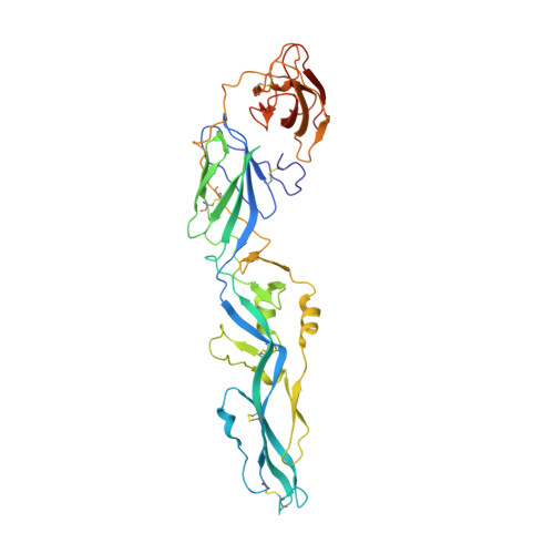
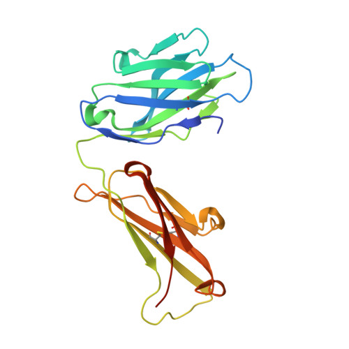
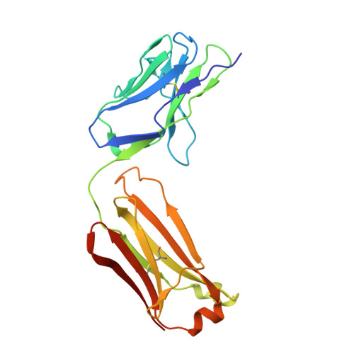
5JHL

Crystal structure of zika virus envelope protein in complex with a flavivirus broadly-protective antibody


Experimental Data Snapshot

  • Method: X-RAY DIFFRACTION
  • Resolution: 3.00 Å
  • R-Value Free: 0.269 
  • R-Value Work: 0.228 
  • R-Value Observed: 0.230 

Starting Model: experimental
View more details

wwPDB Validation   3D Report Full Report


This is version 1.4 of the entry. See complete history


Literature

Structures of the Zika Virus Envelope Protein and Its Complex with a Flavivirus Broadly Protective Antibody.

Dai, L.Song, J.Lu, X.Deng, Y.Q.Musyoki, A.M.Cheng, H.Zhang, Y.Yuan, Y.Song, H.Haywood, J.Xiao, H.Yan, J.Shi, Y.Qin, C.F.Qi, J.Gao, G.F.

(2016) Cell Host Microbe 19: 696-704

  • DOI: https://doi.org/10.1016/j.chom.2016.04.013
  • Primary Citation of Related Structures:  
    5JHL, 5JHM

  • PubMed Abstract: 

    Zika virus (ZIKV), a mosquito-borne flavivirus, is a current global public health concern. The flavivirus envelope (E) glycoprotein is responsible for virus entry and represents a major target of neutralizing antibodies for other flaviviruses. Here, we report the structures of ZIKV E protein at 2.0 Å and in complex with a flavivirus broadly neutralizing murine antibody 2A10G6 at 3.0 Å. ZIKV-E resembles all the known flavivirus E structures but contains a unique, positively charged patch adjacent to the fusion loop region of the juxtaposed monomer, which may influence host attachment. The ZIKV-E-2A10G6 complex structure reveals antibody recognition of a highly conserved fusion loop. 2A10G6 binds to ZIKV-E with high affinity in vitro and neutralizes currently circulating ZIKV strains in vitro and in mice. The E protein fusion loop epitope represents a potential candidate for therapeutic antibodies against ZIKV.


  • Organizational Affiliation

    Research Network of Immunity and Health (RNIH), Beijing Institutes of Life Science, Chinese Academy of Sciences, Beijing 100101, China.


Macromolecules
Find similar proteins by:  (by identity cutoff)  |  3D Structure
Entity ID: 1
MoleculeChains Sequence LengthOrganismDetailsImage
envelope protein416Zika virusMutation(s): 0 
Membrane Entity: Yes 
UniProt
Find proteins for A0A0X9QZM7 (Zika virus)
Explore A0A0X9QZM7 
Go to UniProtKB:  A0A0X9QZM7
Entity Groups  
Sequence Clusters30% Identity50% Identity70% Identity90% Identity95% Identity100% Identity
UniProt GroupA0A0X9QZM7
Sequence Annotations
Expand
  • Reference Sequence
Find similar proteins by:  (by identity cutoff)  |  3D Structure
Entity ID: 2
MoleculeChains Sequence LengthOrganismDetailsImage
Antibody Heavy chainB [auth H]231Mus musculusMutation(s): 0 
Entity Groups  
Sequence Clusters30% Identity50% Identity70% Identity90% Identity95% Identity100% Identity
Sequence Annotations
Expand
  • Reference Sequence
Find similar proteins by:  (by identity cutoff)  |  3D Structure
Entity ID: 3
MoleculeChains Sequence LengthOrganismDetailsImage
antibody Light chainC [auth L]216Mus musculusMutation(s): 0 
Entity Groups  
Sequence Clusters30% Identity50% Identity70% Identity90% Identity95% Identity100% Identity
Sequence Annotations
Expand
  • Reference Sequence
Experimental Data & Validation

Experimental Data

  • Method: X-RAY DIFFRACTION
  • Resolution: 3.00 Å
  • R-Value Free: 0.269 
  • R-Value Work: 0.228 
  • R-Value Observed: 0.230 
  • Space Group: P 1 21 1
Unit Cell:
Length ( Å )Angle ( ˚ )
a = 49.6α = 90
b = 104.856β = 102.7
c = 81.862γ = 90
Software Package:
Software NamePurpose
PHENIXrefinement
HKL-2000data reduction
HKL-2000data scaling
PHASERphasing

Structure Validation

View Full Validation Report


Entry History 

Deposition Data

Revision History  (Full details and data files)

  • Version 1.0: 2016-05-11
    Type: Initial release
  • Version 1.1: 2016-05-18
    Changes: Database references
  • Version 1.2: 2016-08-10
    Changes: Source and taxonomy
  • Version 1.3: 2023-11-08
    Changes: Data collection, Database references, Derived calculations, Refinement description
  • Version 1.4: 2024-11-06
    Changes: Structure summary