RCSB PDB - 5IKV: The Structure of Flufenamic Acid Bound to Human Cyclooxygenase-2

 5IKV

The Structure of Flufenamic Acid Bound to Human Cyclooxygenase-2


Experimental Data Snapshot

  • Method: X-RAY DIFFRACTION
  • Resolution: 2.51 Å
  • R-Value Free: 0.224 
  • R-Value Work: 0.185 
  • R-Value Observed: 0.187 

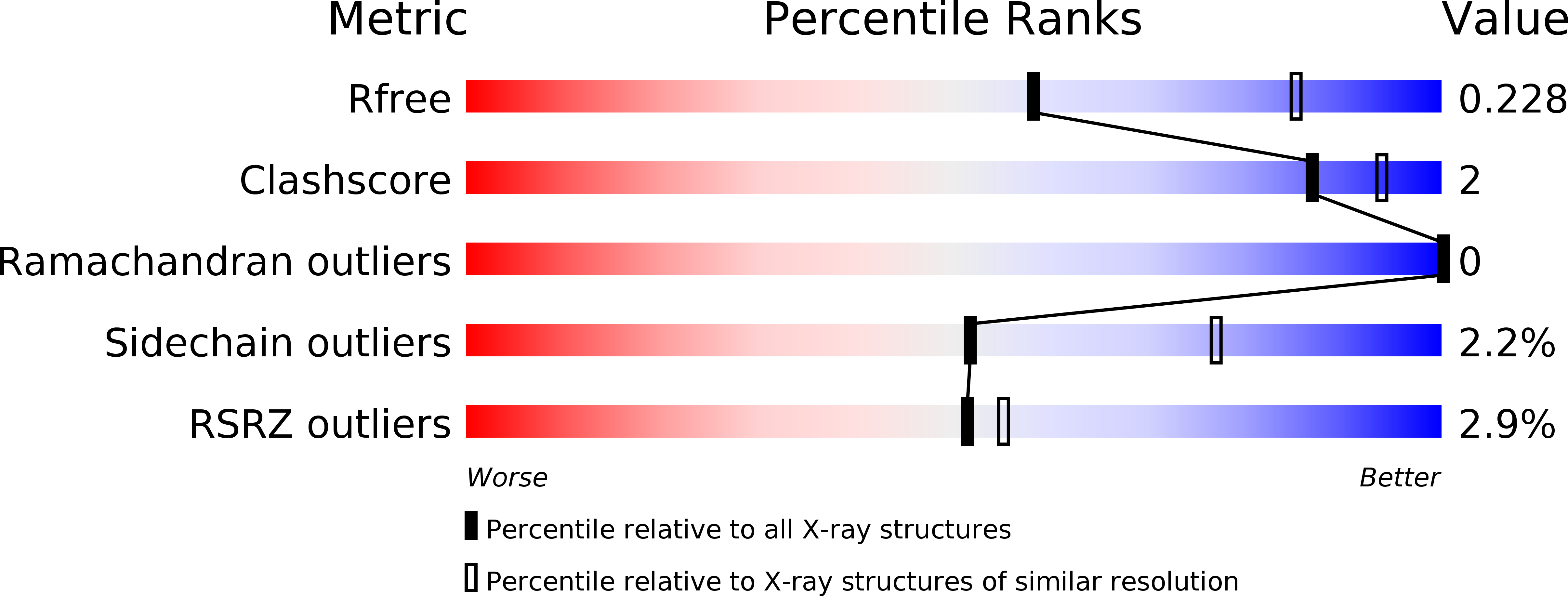
wwPDB Validation   3D Report Full Report


Ligand Structure Quality Assessment 

Created with Raphaël 2.3.0Worse 01 BetterLigand structure goodness of fit to experimental dataBest fitted COHClick on this verticalbar to view detailsBest fitted BOGClick on this verticalbar to view detailsBest fitted FLFClick on this verticalbar to view detailsBest fitted NAGClick on this verticalbar to view details

This is version 2.1 of the entry. See complete history


Literature

Substrate-selective Inhibition of Cyclooxygeanse-2 by Fenamic Acid Derivatives Is Dependent on Peroxide Tone.

Orlando, B.J.Malkowski, M.G.

(2016) J Biol Chem 291: 15069-15081

  • DOI: https://doi.org/10.1074/jbc.M116.725713
  • Primary Citation of Related Structures:  
    5IKQ, 5IKR, 5IKT, 5IKV

  • PubMed Abstract: 

    Cyclooxygenase-2 (COX-2) catalyzes the oxygenation of arachidonic acid (AA) and endocannabinoid substrates, placing the enzyme at a unique junction between the eicosanoid and endocannabinoid signaling pathways. COX-2 is a sequence homodimer, but the enzyme displays half-of-site reactivity, such that only one monomer of the dimer is active at a given time. Certain rapid reversible, competitive nonsteroidal anti-inflammatory drugs (NSAIDs) have been shown to inhibit COX-2 in a substrate-selective manner, with the binding of inhibitor to a single monomer sufficient to inhibit the oxygenation of endocannabinoids but not arachidonic acid. The underlying mechanism responsible for substrate-selective inhibition has remained elusive. We utilized structural and biophysical methods to evaluate flufenamic acid, meclofenamic acid, mefenamic acid, and tolfenamic acid for their ability to act as substrate-selective inhibitors. Crystal structures of each drug in complex with human COX-2 revealed that the inhibitor binds within the cyclooxygenase channel in an inverted orientation, with the carboxylate group interacting with Tyr-385 and Ser-530 at the top of the channel. Tryptophan fluorescence quenching, continuous-wave electron spin resonance, and UV-visible spectroscopy demonstrate that flufenamic acid, mefenamic acid, and tolfenamic acid are substrate-selective inhibitors that bind rapidly to COX-2, quench tyrosyl radicals, and reduce higher oxidation states of the heme moiety. Substrate-selective inhibition was attenuated by the addition of the lipid peroxide 15-hydroperoxyeicosatertaenoic acid. Collectively, these studies implicate peroxide tone as an important mechanistic component of substrate-selective inhibition by flufenamic acid, mefenamic acid, and tolfenamic acid.


  • Organizational Affiliation

    From the Department of Structural Biology, The State University of New York at Buffalo and.


Macromolecules
Find similar proteins by:  (by identity cutoff)  |  3D Structure
Entity ID: 1
MoleculeChains Sequence LengthOrganismDetailsImage
Prostaglandin G/H synthase 2
A, B
551Homo sapiensMutation(s): 0 
Gene Names: PTGS2COX2
EC: 1.14.99.1
UniProt & NIH Common Fund Data Resources
Find proteins for P35354 (Homo sapiens)
Explore P35354 
Go to UniProtKB:  P35354
PHAROS:  P35354
GTEx:  ENSG00000073756 
Entity Groups  
Sequence Clusters30% Identity50% Identity70% Identity90% Identity95% Identity100% Identity
UniProt GroupP35354
Glycosylation
Glycosylation Sites: 2Go to GlyGen: P35354-1
Sequence Annotations
Expand
  • Reference Sequence
Oligosaccharides

Help

Entity ID: 2
MoleculeChains Length2D Diagram Glycosylation3D Interactions
alpha-D-mannopyranose-(1-4)-2-acetamido-2-deoxy-beta-D-glucopyranose-(1-4)-2-acetamido-2-deoxy-beta-D-glucopyranose
C, D
3N-Glycosylation
Glycosylation Resources
GlyTouCan:  G62182OO
GlyCosmos:  G62182OO
GlyGen:  G62182OO
Small Molecules
Ligands 5 Unique
IDChains Name / Formula / InChI Key2D Diagram3D Interactions
COH
Query on COH

Download Ideal Coordinates CCD File 
F [auth A],
O [auth B]
PROTOPORPHYRIN IX CONTAINING CO
C34 H32 Co N4 O4
AQTFKGDWFRRIHR-RGGAHWMASA-L
BOG
Query on BOG

Download Ideal Coordinates CCD File 
M [auth A]octyl beta-D-glucopyranoside
C14 H28 O6
HEGSGKPQLMEBJL-RKQHYHRCSA-N
FLF
Query on FLF

Download Ideal Coordinates CCD File 
E [auth A],
N [auth B]
2-[[3-(TRIFLUOROMETHYL)PHENYL]AMINO] BENZOIC ACID
C14 H10 F3 N O2
LPEPZBJOKDYZAD-UHFFFAOYSA-N
NAG
Query on NAG

Download Ideal Coordinates CCD File 
G [auth A],
P [auth B]
2-acetamido-2-deoxy-beta-D-glucopyranose
C8 H15 N O6
OVRNDRQMDRJTHS-FMDGEEDCSA-N
NH4
Query on NH4

Download Ideal Coordinates CCD File 
H [auth A]
I [auth A]
J [auth A]
K [auth A]
L [auth A]
AMMONIUM ION
H4 N
QGZKDVFQNNGYKY-UHFFFAOYSA-O
Binding Affinity Annotations 
IDSourceBinding Affinity
FLF BindingDB:  5IKV IC50: min: 16, max: 9300 (nM) from 3 assay(s)
Experimental Data & Validation

Experimental Data

  • Method: X-RAY DIFFRACTION
  • Resolution: 2.51 Å
  • R-Value Free: 0.224 
  • R-Value Work: 0.185 
  • R-Value Observed: 0.187 
  • Space Group: I 2 2 2
Unit Cell:
Length ( Å )Angle ( ˚ )
a = 126.918α = 90
b = 149.334β = 90
c = 184.766γ = 90
Software Package:
Software NamePurpose
PHENIXrefinement
HKL-2000data reduction
HKL-2000data scaling
PHASERphasing

Structure Validation

View Full Validation Report


Ligand Structure Quality Assessment 

Created with Raphaël 2.3.0Worse 01 BetterLigand structure goodness of fit to experimental dataBest fitted COHClick on this verticalbar to view detailsBest fitted BOGClick on this verticalbar to view detailsBest fitted FLFClick on this verticalbar to view detailsBest fitted NAGClick on this verticalbar to view details

Entry History & Funding Information

Deposition Data


Funding OrganizationLocationGrant Number
National Institutes of Health/National Institute of General Medical Sciences (NIH/NIGMS)United StatesGM115386

Revision History  (Full details and data files)

  • Version 1.0: 2016-05-25
    Type: Initial release
  • Version 1.1: 2016-06-08
    Changes: Data collection, Database references
  • Version 1.2: 2016-07-27
    Changes: Database references
  • Version 1.3: 2017-09-27
    Changes: Advisory, Author supporting evidence, Database references, Derived calculations
  • Version 1.4: 2019-12-25
    Changes: Author supporting evidence
  • Version 2.0: 2020-07-29
    Type: Remediation
    Reason: Carbohydrate remediation
    Changes: Atomic model, Data collection, Derived calculations, Structure summary
  • Version 2.1: 2024-10-23
    Changes: Advisory, Data collection, Database references, Structure summary