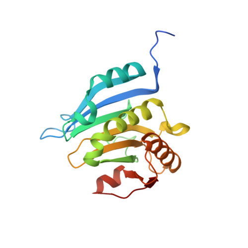
5GW6

Water-Bridge Mediates Recognition of mRNA Cap in eIF4E


Experimental Data Snapshot

  • Method: X-RAY DIFFRACTION
  • Resolution: 1.97 Å
  • R-Value Free: 0.228 
  • R-Value Work: 0.197 
  • R-Value Observed: 0.198 

Starting Model: experimental
View more details

wwPDB Validation   3D Report Full Report


This is version 1.2 of the entry. See complete history


Literature

Water-Bridge Mediates Recognition of mRNA Cap in eIF4E

Lama, D.Pradhan, M.R.Brown, C.J.Eapen, R.S.Joseph, T.L.Kwoh, C.K.Lane, D.P.Verma, C.S.

(2017) Structure 25: 188-194

  • DOI: https://doi.org/10.1016/j.str.2016.11.006
  • Primary Citation of Related Structures:  
    5GW6

  • PubMed Abstract: 

    Ligand binding pockets in proteins contain water molecules, which play important roles in modulating protein-ligand interactions. Available crystallographic data for the 5' mRNA cap-binding pocket of the translation initiation factor protein eIF4E shows several structurally conserved waters, which also persist in molecular dynamics simulations. These waters engage an intricate hydrogen-bond network between the cap and protein. Two crystallographic waters in the cleft of the pocket show a high degree of conservation and bridge two residues, which are part of an evolutionarily conserved scaffold. This appears to be a preformed recognition module for the cap with the two structural waters facilitating an efficient interaction. This is also recapitulated in a new crystal structure of the apo protein. These findings open new windows for the design and screening of compounds targeting eIF4E.


  • Organizational Affiliation

    Bioinformatics Institute, A(∗)STAR (Agency for Science, Technology and Research), 30 Biopolis Street, #07-01 Matrix, Singapore 138671, Singapore. Electronic address: [email protected].


Macromolecules
Find similar proteins by:  (by identity cutoff)  |  3D Structure
Entity ID: 1
MoleculeChains Sequence LengthOrganismDetailsImage
Eukaryotic translation initiation factor 4E196Homo sapiensMutation(s): 3 
Gene Names: EIF4EEIF4EL1EIF4F
UniProt & NIH Common Fund Data Resources
Find proteins for P06730 (Homo sapiens)
Explore P06730 
Go to UniProtKB:  P06730
PHAROS:  P06730
GTEx:  ENSG00000151247 
Entity Groups  
Sequence Clusters30% Identity50% Identity70% Identity90% Identity95% Identity100% Identity
UniProt GroupP06730
Sequence Annotations
Expand
  • Reference Sequence
Experimental Data & Validation

Experimental Data

  • Method: X-RAY DIFFRACTION
  • Resolution: 1.97 Å
  • R-Value Free: 0.228 
  • R-Value Work: 0.197 
  • R-Value Observed: 0.198 
  • Space Group: C 2 2 21
Unit Cell:
Length ( Å )Angle ( ˚ )
a = 121.695α = 90
b = 128.024β = 90
c = 38.469γ = 90
Software Package:
Software NamePurpose
REFMACrefinement
HKL-2000data reduction
HKL-2000data scaling
PHASERphasing

Structure Validation

View Full Validation Report


Entry History 

Deposition Data

  • Released Date: 2016-10-19 
  • Deposition Author(s): Brown, C.J.
  • This entry supersedes: 5ABI

Revision History  (Full details and data files)

  • Version 1.0: 2016-10-19
    Type: Initial release
  • Version 1.1: 2017-02-01
    Changes: Database references
  • Version 1.2: 2023-11-08
    Changes: Data collection, Database references, Refinement description