5G1A

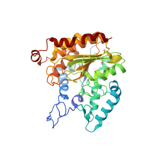
Bordetella Alcaligenes HDAH bound to PFSAHA


Experimental Data Snapshot

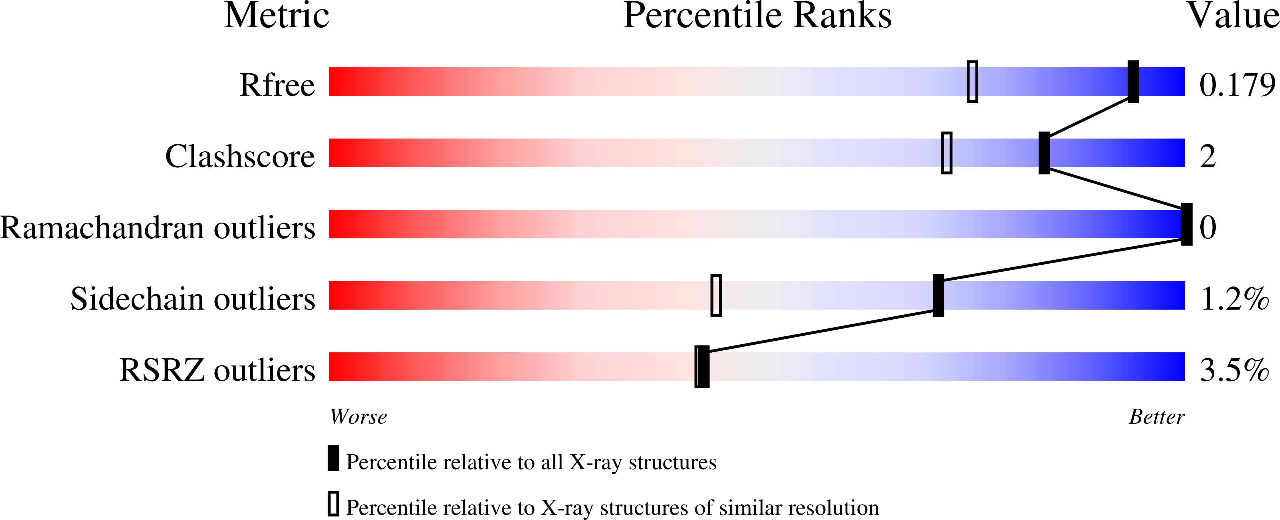
  • Method: X-RAY DIFFRACTION
  • Resolution: 1.42 Å
  • R-Value Free: 0.170 
  • R-Value Work: 0.143 
  • R-Value Observed: 0.145 

Starting Model: experimental
View more details

wwPDB Validation   3D Report Full Report


Ligand Structure Quality Assessment 


This is version 1.4 of the entry. See complete history


Literature

The thermodynamic signature of ligand binding to histone deacetylase-like amidohydrolases is most sensitive to the flexibility in the L2-loop lining the active site pocket.

Meyners, C.Kramer, A.Yildiz, O.Meyer-Almes, F.J.

(2017) Biochim Biophys Acta 1861: 1855-1863

  • DOI: https://doi.org/10.1016/j.bbagen.2017.04.001
  • Primary Citation of Related Structures:  
    5G17, 5G1A, 5G1B

  • PubMed Abstract: 

    The analysis of the thermodynamic driving forces of ligand-protein binding has been suggested to be a key component for the selection and optimization of active compounds into drug candidates. The binding enthalpy as deduced from isothermal titration calorimetry (ITC) is usually interpreted assuming single-step binding of a ligand to one conformation of the target protein. Although successful in many cases, these assumptions are oversimplified approximations of the reality with flexible proteins and complicated binding mechanism in many if not most cases. The relationship between protein flexibility and thermodynamic signature of ligand binding is largely understudied. Directed mutagenesis, X-ray crystallography, enzyme kinetics and ITC methods were combined to dissect the influence of loop flexibility on the thermodynamics and mechanism of ligand binding to histone deacetylase (HDAC)-like amidohydrolases. The general ligand-protein binding mechanism comprises an energetically demanding gate opening step followed by physical binding. Increased flexibility of the L2-loop in HDAC-like amidohydrolases facilitates access of ligands to the binding pocket resulting in predominantly enthalpy-driven complex formation. The study provides evidence for the great importance of flexibility adjacent to the active site channel for the mechanism and observed thermodynamic driving forces of molecular recognition in HDAC like enzymes. The flexibility or malleability in regions adjacent to binding pockets should be given more attention when designing better drug candidates. The presented case study also suggests that the observed binding enthalpy of protein-ligand systems should be interpreted with caution, since more complicated binding mechanisms may obscure the significance regarding potential drug likeness.


  • Organizational Affiliation

    Department of Chemical Engineering and Biotechnology, University of Applied Sciences Darmstadt, Haardtring 100, 64295 Darmstadt, Germany.


Macromolecules
Find similar proteins by:  (by identity cutoff)  |  3D Structure
Entity ID: 1
MoleculeChains Sequence LengthOrganismDetailsImage
HISTONE DEACETYLASE-LIKE AMIDOHYDROLASE
A, B
371AlcaligenesMutation(s): 0 
EC: 3.5.1.4 (PDB Primary Data), 3.5.1 (UniProt)
UniProt
Find proteins for Q70I53 (Alcaligenes sp. (strain DSM 11172))
Explore Q70I53 
Go to UniProtKB:  Q70I53
Entity Groups  
Sequence Clusters30% Identity50% Identity70% Identity90% Identity95% Identity100% Identity
UniProt GroupQ70I53
Sequence Annotations
Expand
  • Reference Sequence
Small Molecules
Ligands 5 Unique
IDChains Name / Formula / InChI Key2D Diagram3D Interactions
7H1
Query on 7H1

Download Ideal Coordinates CCD File 
F [auth A],
P [auth B]
2,2,3,3,4,4,5,5,6,6,7,7-dodecakis(fluoranyl)-~{N}-oxidanyl-~{N}'-phenyl-octanediamide
C14 H8 F12 N2 O3
PVEUUUJZCFCCIZ-UHFFFAOYSA-N
1PE
Query on 1PE

Download Ideal Coordinates CCD File 
G [auth A],
Q [auth B]
PENTAETHYLENE GLYCOL
C10 H22 O6
JLFNLZLINWHATN-UHFFFAOYSA-N
PEG
Query on PEG

Download Ideal Coordinates CCD File 
H [auth A]
I [auth A]
J [auth A]
K [auth A]
L [auth A]
H [auth A],
I [auth A],
J [auth A],
K [auth A],
L [auth A],
R [auth B],
S [auth B],
T [auth B]
DI(HYDROXYETHYL)ETHER
C4 H10 O3
MTHSVFCYNBDYFN-UHFFFAOYSA-N
ZN
Query on ZN

Download Ideal Coordinates CCD File 
C [auth A],
M [auth B]
ZINC ION
Zn
PTFCDOFLOPIGGS-UHFFFAOYSA-N
K
Query on K

Download Ideal Coordinates CCD File 
D [auth A],
E [auth A],
N [auth B],
O [auth B]
POTASSIUM ION
K
NPYPAHLBTDXSSS-UHFFFAOYSA-N
Binding Affinity Annotations 
IDSourceBinding Affinity
7H1 BindingDB:  5G1A Kd: 540 (nM) from 1 assay(s)
Experimental Data & Validation

Experimental Data

  • Method: X-RAY DIFFRACTION
  • Resolution: 1.42 Å
  • R-Value Free: 0.170 
  • R-Value Work: 0.143 
  • R-Value Observed: 0.145 
  • Space Group: P 42 21 2
Unit Cell:
Length ( Å )Angle ( ˚ )
a = 101.44α = 90
b = 101.44β = 90
c = 175.93γ = 90
Software Package:
Software NamePurpose
REFMACrefinement
MOSFLMdata reduction
Aimlessdata scaling
MOLREPphasing

Structure Validation

View Full Validation Report


Ligand Structure Quality Assessment 


Entry History 

Deposition Data

Revision History  (Full details and data files)

  • Version 1.0: 2017-04-12
    Type: Initial release
  • Version 1.1: 2017-04-19
    Changes: Database references
  • Version 1.2: 2017-07-05
    Changes: Database references
  • Version 1.3: 2017-08-23
    Changes: Data collection
  • Version 1.4: 2024-01-10
    Changes: Data collection, Database references, Derived calculations, Other, Refinement description