5CEI | pdb_00005cei

Crystal structure of CDK8:Cyclin C complex with compound 22


Experimental Data Snapshot

  • Method: X-RAY DIFFRACTION
  • Resolution: 2.24 Å
  • R-Value Free: 
    0.227 (Depositor), 0.230 (DCC) 
  • R-Value Work: 
    0.173 (Depositor), 0.180 (DCC) 
  • R-Value Observed: 
    0.175 (Depositor) 

wwPDB Validation   3D Report Full Report


Ligand Structure Quality Assessment 

Created with Raphaël 2.3.0Worse 01 BetterLigand structure goodness of fit to experimental dataBest fitted 50RClick on this verticalbar to view details

This is version 1.2 of the entry. See complete history


Literature

Development of a Potent, Specific CDK8 Kinase Inhibitor Which Phenocopies CDK8/19 Knockout Cells.

Koehler, M.F.Bergeron, P.Blackwood, E.M.Bowman, K.Clark, K.R.Firestein, R.Kiefer, J.R.Maskos, K.McCleland, M.L.Orren, L.Salphati, L.Schmidt, S.Schneider, E.V.Wu, J.Beresini, M.H.

(2016) ACS Med Chem Lett 7: 223-228

  • DOI: https://doi.org/10.1021/acsmedchemlett.5b00278
  • Primary Citation of Related Structures:  
    5CEI

  • PubMed Abstract: 

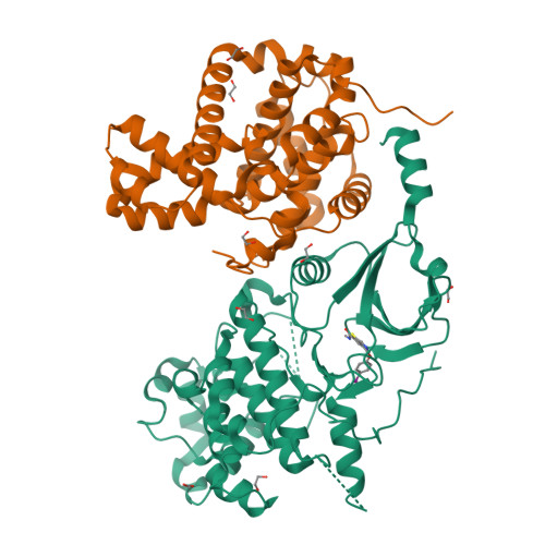
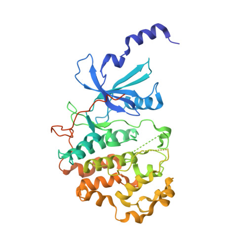
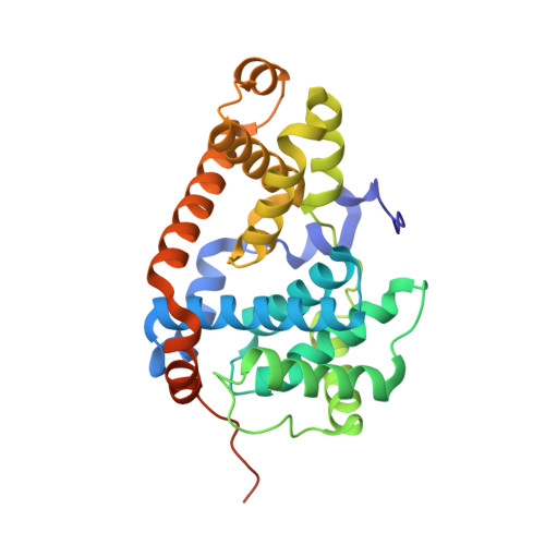
    Beginning with promiscuous COT inhibitors, which were found to inhibit CDK8, a series of 6-aza-benzothiophene containing compounds were developed into potent, selective CDK8 inhibitors. When cocrystallized with CDK8 and cyclin C, these compounds exhibit an unusual binding mode, making a single hydrogen bond to the hinge residue A100, a second to K252, and a key cation-π interaction with R356. Structure-based drug design resulted in tool compounds 13 and 32, which are highly potent, kinase selective, permeable compounds with a free fraction >2% and no measurable efflux. Despite these attractive properties, these compounds exhibit weak antiproliferative activity in the HCT-116 colon cancer cell line. Further examination of the activity of 32 in this cell line revealed that the compound reduced phosphorylation of the known CDK8 substrate STAT1 in a manner identical to a CDK8 knockout clone, illustrating the complex effects of inhibition of CDK8 kinase activity in proliferation in these cells.


  • Organizational Affiliation

    Departments of Discovery Chemistry, Translational Oncology, Structural Biology, Biochemical and Cellular Pharmacology, Pathology, Drug Metabolism and Pharmacokinetics, and Structural Biology, Genentech, Inc. , 1 DNA Way, South San Francisco, California 94080, United States.


Macromolecules
Find similar proteins by:  (by identity cutoff)  |  3D Structure
Entity ID: 1
MoleculeChains Sequence LengthOrganismDetailsImage
Cyclin-dependent kinase 8406Homo sapiensMutation(s): 0 
Gene Names: CDK8
EC: 2.7.11.22 (PDB Primary Data), 2.7.11.23 (PDB Primary Data)
UniProt & NIH Common Fund Data Resources
Find proteins for P49336 (Homo sapiens)
Explore P49336 
Go to UniProtKB:  P49336
PHAROS:  P49336
GTEx:  ENSG00000132964 
Entity Groups  
Sequence Clusters30% Identity50% Identity70% Identity90% Identity95% Identity100% Identity
UniProt GroupP49336
Sequence Annotations
Expand
  • Reference Sequence
Find similar proteins by:  (by identity cutoff)  |  3D Structure
Entity ID: 2
MoleculeChains Sequence LengthOrganismDetailsImage
Cyclin-C287Homo sapiensMutation(s): 0 
Gene Names: CCNC
UniProt & NIH Common Fund Data Resources
Find proteins for P24863 (Homo sapiens)
Explore P24863 
Go to UniProtKB:  P24863
PHAROS:  P24863
GTEx:  ENSG00000112237 
Entity Groups  
Sequence Clusters30% Identity50% Identity70% Identity90% Identity95% Identity100% Identity
UniProt GroupP24863
Sequence Annotations
Expand
  • Reference Sequence
Small Molecules
Ligands 3 Unique
IDChains Name / Formula / InChI Key2D Diagram3D Interactions
50R
Query on 50R

Download Ideal Coordinates CCD File 
J [auth A]4-(4-iodophenoxy)-N-methylthieno[2,3-c]pyridine-2-carboxamide
C15 H11 I N2 O2 S
QHMOZKVAGIEXJR-UHFFFAOYSA-N
EDO
Query on EDO

Download Ideal Coordinates CCD File 
C [auth A]
D [auth A]
E [auth A]
F [auth A]
K [auth A]
1,2-ETHANEDIOL
C2 H6 O2
LYCAIKOWRPUZTN-UHFFFAOYSA-N
FMT
Query on FMT

Download Ideal Coordinates CCD File 
G [auth A]
H [auth A]
I [auth A]
N [auth B]
O [auth B]
FORMIC ACID
C H2 O2
BDAGIHXWWSANSR-UHFFFAOYSA-N
Binding Affinity Annotations 
IDSourceBinding Affinity
50R BindingDB:  5CEI IC50: 5.3 (nM) from 1 assay(s)
Experimental Data & Validation

Experimental Data

  • Method: X-RAY DIFFRACTION
  • Resolution: 2.24 Å
  • R-Value Free:  0.227 (Depositor), 0.230 (DCC) 
  • R-Value Work:  0.173 (Depositor), 0.180 (DCC) 
  • R-Value Observed: 0.175 (Depositor) 
Space Group: P 21 21 21
Unit Cell:
Length ( Å )Angle ( ˚ )
a = 71.128α = 90
b = 70.09β = 90
c = 167.448γ = 90
Software Package:
Software NamePurpose
PHENIXrefinement
SCALAdata scaling
PDB_EXTRACTdata extraction
XDSdata reduction
PHASERphasing

Structure Validation

View Full Validation Report


Ligand Structure Quality Assessment 

Created with Raphaël 2.3.0Worse 01 BetterLigand structure goodness of fit to experimental dataBest fitted 50RClick on this verticalbar to view details

Entry History 

Deposition Data

Revision History  (Full details and data files)

  • Version 1.0: 2016-02-10
    Type: Initial release
  • Version 1.1: 2016-05-11
    Changes: Database references
  • Version 1.2: 2024-03-06
    Changes: Data collection, Database references, Derived calculations