4GUP | pdb_00004gup

Structure of MHC-class I related molecule MR1


Experimental Data Snapshot

  • Method: X-RAY DIFFRACTION
  • Resolution: 3.20 Å
  • R-Value Free: 
    0.260 (Depositor), 0.290 (DCC) 
  • R-Value Work: 
    0.193 (Depositor), 0.210 (DCC) 
  • R-Value Observed: 
    0.196 (Depositor) 

Starting Model: experimental
View more details

wwPDB Validation   3D Report Full Report


Ligand Structure Quality Assessment 

Created with Raphaël 2.3.0Worse 01 BetterLigand structure goodness of fit to experimental dataBest fitted 6FPClick on this verticalbar to view details

This is version 1.4 of the entry. See complete history


Literature

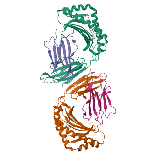
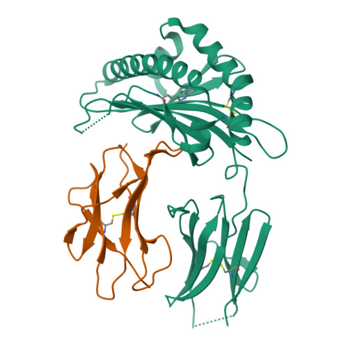
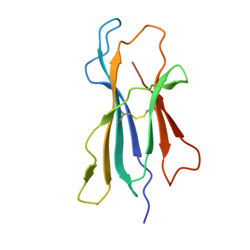
MR1 presents microbial vitamin B metabolites to MAIT cells

Kjer-Nielsen, L.Patel, O.Corbett, A.J.Le Nours, J.Meehan, B.Liu, L.Bhati, M.Chen, Z.Kostenko, L.Reantragoon, R.Williamson, N.A.Purcell, A.W.Dudek, N.L.McConville, M.J.O'Hair, R.A.J.Khairallah, G.N.Godfrey, D.I.Fairlie, D.P.Rossjohn, J.McCluskey, J.

(2012) Nature 491: 717-723

  • DOI: https://doi.org/10.1038/nature11605
  • Primary Citation of Related Structures:  
    4GUP

  • PubMed Abstract: 

    Antigen-presenting molecules, encoded by the major histocompatibility complex (MHC) and CD1 family, bind peptide- and lipid-based antigens, respectively, for recognition by T cells. Mucosal-associated invariant T (MAIT) cells are an abundant population of innate-like T cells in humans that are activated by an antigen(s) bound to the MHC class I-like molecule MR1. Although the identity of MR1-restricted antigen(s) is unknown, it is present in numerous bacteria and yeast. Here we show that the structure and chemistry within the antigen-binding cleft of MR1 is distinct from the MHC and CD1 families. MR1 is ideally suited to bind ligands originating from vitamin metabolites. The structure of MR1 in complex with 6-formyl pterin, a folic acid (vitamin B9) metabolite, shows the pterin ring sequestered within MR1. Furthermore, we characterize related MR1-restricted vitamin derivatives, originating from the bacterial riboflavin (vitamin B2) biosynthetic pathway, which specifically and potently activate MAIT cells. Accordingly, we show that metabolites of vitamin B represent a class of antigen that are presented by MR1 for MAIT-cell immunosurveillance. As many vitamin biosynthetic pathways are unique to bacteria and yeast, our data suggest that MAIT cells use these metabolites to detect microbial infection.


  • Organizational Affiliation

    Department of Microbiology & Immunology, University of Melbourne, Parkville, Victoria 3010, Australia.


Macromolecules
Find similar proteins by:  (by identity cutoff)  |  3D Structure
Entity ID: 1
MoleculeChains Sequence LengthOrganismDetailsImage
Major histocompatibility complex class I-related gene protein
A, C
271Homo sapiensMutation(s): 1 
Gene Names: MR1
UniProt & NIH Common Fund Data Resources
Find proteins for Q95460 (Homo sapiens)
Explore Q95460 
Go to UniProtKB:  Q95460
PHAROS:  Q95460
GTEx:  ENSG00000153029 
Entity Groups  
Sequence Clusters30% Identity50% Identity70% Identity90% Identity95% Identity100% Identity
UniProt GroupQ95460
Sequence Annotations
Expand
  • Reference Sequence
Find similar proteins by:  (by identity cutoff)  |  3D Structure
Entity ID: 2
MoleculeChains Sequence LengthOrganismDetailsImage
Beta-2-microglobulin
B, D
99Homo sapiensMutation(s): 0 
Gene Names: B2M
UniProt & NIH Common Fund Data Resources
Find proteins for P61769 (Homo sapiens)
Explore P61769 
Go to UniProtKB:  P61769
PHAROS:  P61769
GTEx:  ENSG00000166710 
Entity Groups  
Sequence Clusters30% Identity50% Identity70% Identity90% Identity95% Identity100% Identity
UniProt GroupP61769
Sequence Annotations
Expand
  • Reference Sequence
Experimental Data & Validation

Experimental Data

  • Method: X-RAY DIFFRACTION
  • Resolution: 3.20 Å
  • R-Value Free:  0.260 (Depositor), 0.290 (DCC) 
  • R-Value Work:  0.193 (Depositor), 0.210 (DCC) 
  • R-Value Observed: 0.196 (Depositor) 
Space Group: P 21 21 21
Unit Cell:
Length ( Å )Angle ( ˚ )
a = 59.154α = 90
b = 89.777β = 90
c = 171.338γ = 90
Software Package:
Software NamePurpose
Blu-Icedata collection
PHASERphasing
BUSTERrefinement
MOSFLMdata reduction
SCALAdata scaling

Structure Validation

View Full Validation Report


Ligand Structure Quality Assessment 

Created with Raphaël 2.3.0Worse 01 BetterLigand structure goodness of fit to experimental dataBest fitted 6FPClick on this verticalbar to view details

Entry History 

Deposition Data

Revision History  (Full details and data files)

  • Version 1.0: 2012-10-17
    Type: Initial release
  • Version 1.1: 2012-10-24
    Changes: Database references
  • Version 1.2: 2012-12-05
    Changes: Database references
  • Version 1.3: 2023-11-08
    Changes: Data collection, Database references, Derived calculations, Refinement description
  • Version 1.4: 2024-10-30
    Changes: Structure summary