4YWT | pdb_00004ywt

Crystal structure of full-length glypican-1 core protein after controlled crystal dehydration to 87% relative humidity


Experimental Data Snapshot

  • Method: X-RAY DIFFRACTION
  • Resolution: 2.38 Å
  • R-Value Free: 
    0.273 (Depositor), 0.270 (DCC) 
  • R-Value Work: 
    0.239 (Depositor), 0.240 (DCC) 
  • R-Value Observed: 
    0.240 (Depositor) 

Starting Model: experimental
View more details

wwPDB Validation   3D Report Full Report


Ligand Structure Quality Assessment 

Created with Raphaël 2.3.0Worse 01 BetterLigand structure goodness of fit to experimental dataBest fitted NAGClick on this verticalbar to view details

This is version 1.7 of the entry. See complete history


Literature

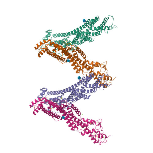
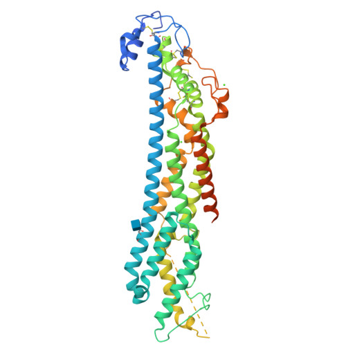
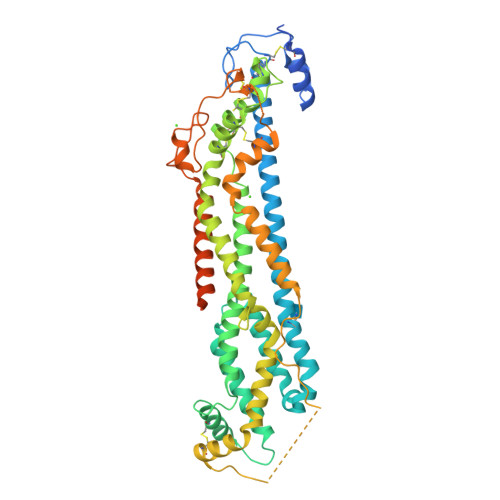
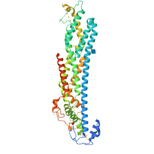
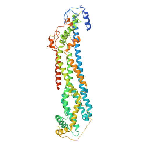
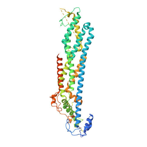
Structural Aspects of N-Glycosylations and the C-terminal Region in Human Glypican-1.

Awad, W.Adamczyk, B.Ornros, J.Karlsson, N.G.Mani, K.Logan, D.T.

(2015) J Biological Chem 290: 22991-23008

  • DOI: https://doi.org/10.1074/jbc.M115.660878
  • Primary Citation of Related Structures:  
    4YWT

  • PubMed Abstract: 

    Glypicans are multifunctional cell surface proteoglycans involved in several important cellular signaling pathways. Glypican-1 (Gpc1) is the predominant heparan sulfate proteoglycan in the developing and adult human brain. The two N-linked glycans and the C-terminal domain that attach the core protein to the cell membrane are not resolved in the Gpc1 crystal structure. Therefore, we have studied Gpc1 using crystallography, small angle x-ray scattering, and chromatographic approaches to elucidate the composition, structure, and function of the N-glycans and the C terminus and also the topology of Gpc1 with respect to the membrane. The C terminus is shown to be highly flexible in solution, but it orients the core protein transverse to the membrane, directing a surface evolutionarily conserved in Gpc1 orthologs toward the membrane, where it may interact with signaling molecules and/or membrane receptors on the cell surface, or even the enzymes involved in heparan sulfate substitution in the Golgi apparatus. Furthermore, the N-glycans are shown to extend the protein stability and lifetime by protection against proteolysis and aggregation.


  • Organizational Affiliation

    From the Department of Biochemistry and Structural Biology, Centre for Molecular Protein Science, Lund University, Box 124, SE-221 00 Lund.


Macromolecules
Find similar proteins by:  (by identity cutoff)  |  3D Structure
Entity ID: 1
MoleculeChains Sequence LengthOrganismDetailsImage
Glypican-1
A, B, C, D
526Homo sapiensMutation(s): 3 
Gene Names: GPC1
UniProt & NIH Common Fund Data Resources
Find proteins for P35052 (Homo sapiens)
Explore P35052 
Go to UniProtKB:  P35052
PHAROS:  P35052
GTEx:  ENSG00000063660 
Entity Groups  
Sequence Clusters30% Identity50% Identity70% Identity90% Identity95% Identity100% Identity
UniProt GroupP35052
Glycosylation
Glycosylation Sites: 1Go to GlyGen: P35052-1
Sequence Annotations
Expand
  • Reference Sequence
Small Molecules
Ligands 2 Unique
IDChains Name / Formula / InChI Key2D Diagram3D Interactions
NAG
Query on NAG

Download Ideal Coordinates CCD File 
E [auth A],
H [auth B],
K [auth C],
O [auth D]
2-acetamido-2-deoxy-beta-D-glucopyranose
C8 H15 N O6
OVRNDRQMDRJTHS-FMDGEEDCSA-N
CA
Query on CA

Download Ideal Coordinates CCD File 
F [auth A]
G [auth A]
I [auth B]
J [auth B]
L [auth C]
CALCIUM ION
Ca
BHPQYMZQTOCNFJ-UHFFFAOYSA-N
Experimental Data & Validation

Experimental Data

  • Method: X-RAY DIFFRACTION
  • Resolution: 2.38 Å
  • R-Value Free:  0.273 (Depositor), 0.270 (DCC) 
  • R-Value Work:  0.239 (Depositor), 0.240 (DCC) 
  • R-Value Observed: 0.240 (Depositor) 
Space Group: P 1 21 1
Unit Cell:
Length ( Å )Angle ( ˚ )
a = 46.78α = 90
b = 166.59β = 90.38
c = 137.7γ = 90
Software Package:
Software NamePurpose
PHENIXrefinement
PDB_EXTRACTdata extraction
XDSdata reduction
XSCALEdata scaling
PHASERphasing

Structure Validation

View Full Validation Report


Ligand Structure Quality Assessment 

Created with Raphaël 2.3.0Worse 01 BetterLigand structure goodness of fit to experimental dataBest fitted NAGClick on this verticalbar to view details

Entry History 

Deposition Data

Revision History  (Full details and data files)

  • Version 1.0: 2015-07-29
    Type: Initial release
  • Version 1.1: 2015-08-05
    Changes: Database references
  • Version 1.2: 2015-09-30
    Changes: Database references
  • Version 1.3: 2018-01-17
    Changes: Data collection
  • Version 1.4: 2019-04-03
    Changes: Data collection, Source and taxonomy
  • Version 1.5: 2020-07-29
    Type: Remediation
    Reason: Carbohydrate remediation
    Changes: Data collection, Derived calculations, Structure summary
  • Version 1.6: 2024-01-10
    Changes: Data collection, Database references, Refinement description, Structure summary
  • Version 1.7: 2024-11-13
    Changes: Structure summary