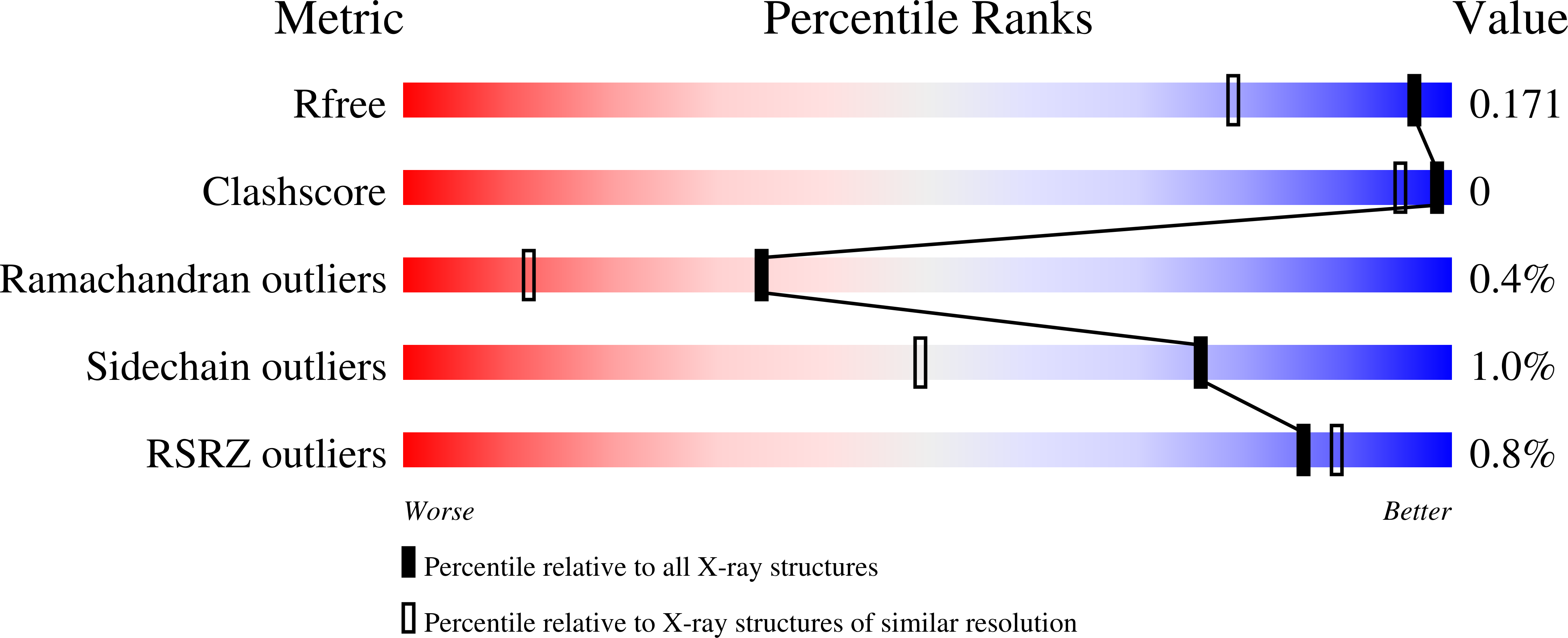
4YP2

Cleavage of nicotinamide adenine dinucleotides by the ribosome inactivating protein from Momordica charantia


Experimental Data Snapshot

  • Method: X-RAY DIFFRACTION
  • Resolution: 1.35 Å
  • R-Value Free: 0.168 
  • R-Value Work: 0.140 
  • R-Value Observed: 0.142 

Starting Model: experimental
View more details

wwPDB Validation   3D Report Full Report


Ligand Structure Quality Assessment 


This is version 1.4 of the entry. See complete history


Literature

Cleavage of nicotinamide adenine dinucleotide by the ribosome-inactivating protein from Momordica charantia.

Vinkovic, M.Dunn, G.Wood, G.E.Husain, J.Wood, S.P.Gill, R.

(2015) Acta Crystallogr F Struct Biol Commun 71: 1152-1155

  • DOI: https://doi.org/10.1107/S2053230X15013540
  • Primary Citation of Related Structures:  
    4YP2, 5CF9

  • PubMed Abstract: 

    The interaction of momordin, a type 1 ribosome-inactivating protein from Momordica charantia, with NADP(+) and NADPH has been investigated by X-ray diffraction analysis of complexes generated by co-crystallization and crystal soaking. It is known that the proteins of this family readily cleave the adenine-ribose bond of adenosine and related nucleotides in the crystal, leaving the product, adenine, bound to the enzyme active site. Surprisingly, the nicotinamide-ribose bond of oxidized NADP(+) is cleaved, leaving nicotinamide bound in the active site in the same position but in a slightly different orientation to that of the five-membered ring of adenine. No binding or cleavage of NADPH was observed at pH 7.4 in these experiments. These observations are in accord with current views of the enzyme mechanism and may contribute to ongoing searches for effective inhibitors.


  • Organizational Affiliation

    Astex Therapeutics, 436 Cambridge Science Park, Milton Road, Cambridge CB4 0QA, England.


Macromolecules
Find similar proteins by:  (by identity cutoff)  |  3D Structure
Entity ID: 1
MoleculeChains Sequence LengthOrganismDetailsImage
Ribosome-inactivating protein momordin IA [auth B]246Momordica charantiaMutation(s): 0 
EC: 3.2.2.22
UniProt
Find proteins for P16094 (Momordica charantia)
Explore P16094 
Go to UniProtKB:  P16094
Entity Groups  
Sequence Clusters30% Identity50% Identity70% Identity90% Identity95% Identity100% Identity
UniProt GroupP16094
Glycosylation
Glycosylation Sites: 1
Sequence Annotations
Expand
  • Reference Sequence
Small Molecules
Ligands 2 Unique
IDChains Name / Formula / InChI Key2D Diagram3D Interactions
NAG
Query on NAG

Download Ideal Coordinates CCD File 
B
2-acetamido-2-deoxy-beta-D-glucopyranose
C8 H15 N O6
OVRNDRQMDRJTHS-FMDGEEDCSA-N
NCA
Query on NCA

Download Ideal Coordinates CCD File 
C [auth B]NICOTINAMIDE
C6 H6 N2 O
DFPAKSUCGFBDDF-UHFFFAOYSA-N
Experimental Data & Validation

Experimental Data

  • Method: X-RAY DIFFRACTION
  • Resolution: 1.35 Å
  • R-Value Free: 0.168 
  • R-Value Work: 0.140 
  • R-Value Observed: 0.142 
  • Space Group: H 3
Unit Cell:
Length ( Å )Angle ( ˚ )
a = 130.1α = 90
b = 130.1β = 90
c = 37.57γ = 120
Software Package:
Software NamePurpose
PHENIXrefinement
xia2data reduction
PDB_EXTRACTdata extraction
Aimlessdata scaling
PHASERphasing

Structure Validation

View Full Validation Report


Ligand Structure Quality Assessment 


Entry History 

Deposition Data

Revision History  (Full details and data files)

  • Version 1.0: 2015-05-20
    Type: Initial release
  • Version 1.1: 2015-09-09
    Changes: Database references
  • Version 1.2: 2020-07-29
    Type: Remediation
    Reason: Carbohydrate remediation
    Changes: Data collection, Derived calculations, Structure summary
  • Version 1.3: 2024-01-10
    Changes: Data collection, Database references, Refinement description, Structure summary
  • Version 1.4: 2024-11-13
    Changes: Structure summary