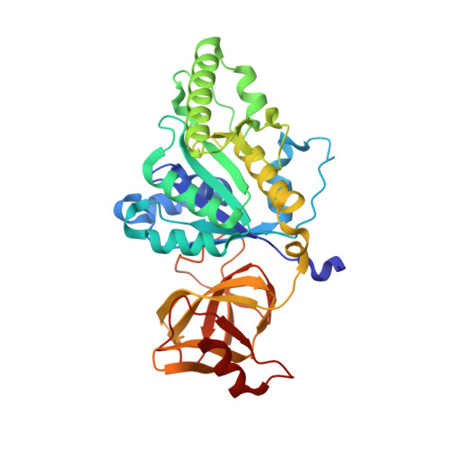
4TMT

Translation initiation factor eIF5B (517-858) mutant D533A from C. thermophilum, bound to GTPgammaS


Experimental Data Snapshot

  • Method: X-RAY DIFFRACTION
  • Resolution: 1.58 Å
  • R-Value Free: 0.194 
  • R-Value Work: 0.169 
  • R-Value Observed: 0.170 

Starting Model: experimental
View more details

wwPDB Validation   3D Report Full Report


Ligand Structure Quality Assessment 


This is version 1.3 of the entry. See complete history


Literature

A monovalent cation acts as structural and catalytic cofactor in translational GTPases.

Kuhle, B.Ficner, R.

(2014) EMBO J 33: 2547-2563

  • DOI: https://doi.org/10.15252/embj.201488517
  • Primary Citation of Related Structures:  
    4TMT, 4TMV, 4TMW, 4TMX, 4TMZ, 4TN1

  • PubMed Abstract: 

    Translational GTPases are universally conserved GTP hydrolyzing enzymes, critical for fidelity and speed of ribosomal protein biosynthesis. Despite their central roles, the mechanisms of GTP-dependent conformational switching and GTP hydrolysis that govern the function of trGTPases remain poorly understood. Here, we provide biochemical and high-resolution structural evidence that eIF5B and aEF1A/EF-Tu bound to GTP or GTPγS coordinate a monovalent cation (M(+)) in their active site. Our data reveal that M(+) ions form constitutive components of the catalytic machinery in trGTPases acting as structural cofactor to stabilize the GTP-bound "on" state. Additionally, the M(+) ion provides a positive charge into the active site analogous to the arginine-finger in the Ras-RasGAP system indicating a similar role as catalytic element that stabilizes the transition state of the hydrolysis reaction. In sequence and structure, the coordination shell for the M(+) ion is, with exception of eIF2γ, highly conserved among trGTPases from bacteria to human. We therefore propose a universal mechanism of M(+)-dependent conformational switching and GTP hydrolysis among trGTPases with important consequences for the interpretation of available biochemical and structural data.


  • Organizational Affiliation

    Abteilung für Molekulare Strukturbiologie, Institut für Mikrobiologie und Genetik Göttinger Zentrum für Molekulare Biowissenschaften Georg-August-Universität Göttingen, Göttingen, Germany [email protected].


Macromolecules
Find similar proteins by:  (by identity cutoff)  |  3D Structure
Entity ID: 1
MoleculeChains Sequence LengthOrganismDetailsImage
eIF5B
A, B
345Thermochaetoides thermophilaMutation(s): 0 
EC: 3.6.5.3
UniProt
Find proteins for G0S8G9 (Chaetomium thermophilum (strain DSM 1495 / CBS 144.50 / IMI 039719))
Explore G0S8G9 
Go to UniProtKB:  G0S8G9
Entity Groups  
Sequence Clusters30% Identity50% Identity70% Identity90% Identity95% Identity100% Identity
UniProt GroupG0S8G9
Sequence Annotations
Expand
  • Reference Sequence
Small Molecules
Ligands 6 Unique
IDChains Name / Formula / InChI Key2D Diagram3D Interactions
GSP
Query on GSP

Download Ideal Coordinates CCD File 
C [auth A],
L [auth B]
5'-GUANOSINE-DIPHOSPHATE-MONOTHIOPHOSPHATE
C10 H16 N5 O13 P3 S
XOFLBQFBSOEHOG-UUOKFMHZSA-N
DTT
Query on DTT

Download Ideal Coordinates CCD File 
K [auth A]2,3-DIHYDROXY-1,4-DITHIOBUTANE
C4 H10 O2 S2
VHJLVAABSRFDPM-IMJSIDKUSA-N
EDO
Query on EDO

Download Ideal Coordinates CCD File 
E [auth A]
F [auth A]
G [auth A]
H [auth A]
I [auth A]
E [auth A],
F [auth A],
G [auth A],
H [auth A],
I [auth A],
N [auth B],
O [auth B],
P [auth B],
Q [auth B],
R [auth B],
S [auth B],
T [auth B]
1,2-ETHANEDIOL
C2 H6 O2
LYCAIKOWRPUZTN-UHFFFAOYSA-N
ACY
Query on ACY

Download Ideal Coordinates CCD File 
U [auth B]ACETIC ACID
C2 H4 O2
QTBSBXVTEAMEQO-UHFFFAOYSA-N
CL
Query on CL

Download Ideal Coordinates CCD File 
J [auth A],
V [auth B]
CHLORIDE ION
Cl
VEXZGXHMUGYJMC-UHFFFAOYSA-M
MG
Query on MG

Download Ideal Coordinates CCD File 
D [auth A],
M [auth B]
MAGNESIUM ION
Mg
JLVVSXFLKOJNIY-UHFFFAOYSA-N
Experimental Data & Validation

Experimental Data

  • Method: X-RAY DIFFRACTION
  • Resolution: 1.58 Å
  • R-Value Free: 0.194 
  • R-Value Work: 0.169 
  • R-Value Observed: 0.170 
  • Space Group: P 1 21 1
Unit Cell:
Length ( Å )Angle ( ˚ )
a = 55.41α = 90
b = 115.91β = 101.37
c = 66.09γ = 90
Software Package:
Software NamePurpose
XDSdata reduction
XSCALEdata scaling
X-PLORmodel building
PDB_EXTRACTdata extraction
PHASERphasing
PHENIXrefinement
XSCALEdata reduction

Structure Validation

View Full Validation Report


Ligand Structure Quality Assessment 


Entry History 

Deposition Data

Revision History  (Full details and data files)

  • Version 1.0: 2014-09-24
    Type: Initial release
  • Version 1.1: 2014-11-19
    Changes: Database references
  • Version 1.2: 2016-02-24
    Changes: Structure summary
  • Version 1.3: 2023-12-20
    Changes: Data collection, Database references, Derived calculations, Refinement description