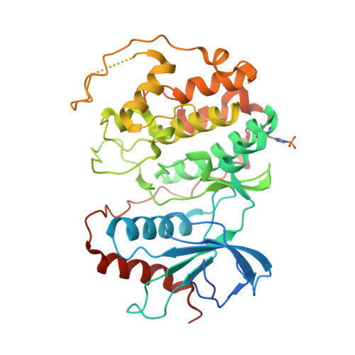
4QPA

Crystal Structure of ERK2 in complex with 7-(1-benzyl-1H-pyrazol-4-yl)-2-(pyridin-4-yl)-5H-pyrrolo[2,3-b]pyrazine


Experimental Data Snapshot

  • Method: X-RAY DIFFRACTION
  • Resolution: 2.85 Å
  • R-Value Free: 0.253 
  • R-Value Work: 0.212 
  • R-Value Observed: 0.214 

wwPDB Validation   3D Report Full Report


Ligand Structure Quality Assessment 


This is version 1.1 of the entry. See complete history


Literature

Fragment-based discovery of potent ERK2 pyrrolopyrazine inhibitors.

Burdick, D.J.Wang, S.Heise, C.Pan, B.Drummond, J.Yin, J.Goeser, L.Magnuson, S.Blaney, J.Moffat, J.Wang, W.Chen, H.

(2015) Bioorg Med Chem Lett 25: 4728-4732

  • DOI: https://doi.org/10.1016/j.bmcl.2015.08.048
  • Primary Citation of Related Structures:  
    4QP1, 4QP2, 4QP3, 4QP4, 4QP6, 4QP7, 4QP8, 4QP9, 4QPA

  • PubMed Abstract: 

    A fragment-based lead discovery approach was used to discover novel ERK2 inhibitors. The crystal structure of N-benzyl-9H-purin-6-amine 1 in complex with ERK2 elucidated its hinge-binding mode. In addition, the simultaneous binding of an imidazole molecule adjacent to 1 suggested a direction for fragment expansion. Structure-based core hopping applied to 1 led to 5H-pyrrolo[3,2-b]pyrazine (3) that afforded direct vectors to probe the pockets of interest while retaining the essential hinge binding elements. Utilizing the new vectors for SAR exploration, the new core 3 was quickly optimized to compound 39 resulting in a greater than 6600-fold improvement in potency.


  • Organizational Affiliation

    Department of Discovery Chemistry, Genentech, Inc., South San Francisco, CA 94080, United States. Electronic address: [email protected].


Macromolecules
Find similar proteins by:  (by identity cutoff)  |  3D Structure
Entity ID: 1
MoleculeChains Sequence LengthOrganismDetailsImage
Mitogen-activated protein kinase 1
A, B
369Homo sapiensMutation(s): 0 
Gene Names: MAPK1ERK2PRKM1PRKM2
EC: 2.7.11.24
UniProt & NIH Common Fund Data Resources
Find proteins for P28482 (Homo sapiens)
Explore P28482 
Go to UniProtKB:  P28482
PHAROS:  P28482
GTEx:  ENSG00000100030 
Entity Groups  
Sequence Clusters30% Identity50% Identity70% Identity90% Identity95% Identity100% Identity
UniProt GroupP28482
Sequence Annotations
Expand
  • Reference Sequence
Small Molecules
Modified Residues  1 Unique
IDChains TypeFormula2D DiagramParent
NEP
Query on NEP
A, B
L-PEPTIDE LINKINGC6 H10 N3 O5 PHIS
Binding Affinity Annotations 
IDSourceBinding Affinity
35W BindingDB:  4QPA IC50: 37 (nM) from 1 assay(s)
Experimental Data & Validation

Experimental Data

  • Method: X-RAY DIFFRACTION
  • Resolution: 2.85 Å
  • R-Value Free: 0.253 
  • R-Value Work: 0.212 
  • R-Value Observed: 0.214 
  • Space Group: P 41 21 2
Unit Cell:
Length ( Å )Angle ( ˚ )
a = 82.703α = 90
b = 82.703β = 90
c = 275.892γ = 90
Software Package:
Software NamePurpose
BOSdata collection
PHASERphasing
PHENIXrefinement
XDSdata reduction
SCALAdata scaling

Structure Validation

View Full Validation Report


Ligand Structure Quality Assessment 


Entry History 

Deposition Data

Revision History  (Full details and data files)

  • Version 1.0: 2015-09-23
    Type: Initial release
  • Version 1.1: 2015-12-16
    Changes: Database references