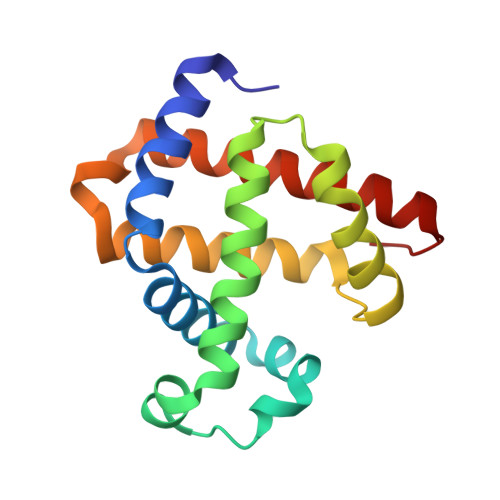
4MXL

X-ray structure of ZnPFeBMb1


Experimental Data Snapshot

  • Method: X-RAY DIFFRACTION
  • Resolution: 1.50 Å
  • R-Value Free: 0.255 
  • R-Value Work: 0.207 
  • R-Value Observed: 0.209 

wwPDB Validation   3D Report Full Report


Ligand Structure Quality Assessment 


This is version 1.3 of the entry. See complete history


Literature

Spectroscopic and computational study of a nonheme iron nitrosyl center in a biosynthetic model of nitric oxide reductase.

Chakraborty, S.Reed, J.Ross, M.Nilges, M.J.Petrik, I.D.Ghosh, S.Hammes-Schiffer, S.Sage, J.T.Zhang, Y.Schulz, C.E.Lu, Y.

(2014) Angew Chem Int Ed Engl 53: 2417-2421

  • DOI: https://doi.org/10.1002/anie.201308431
  • Primary Citation of Related Structures:  
    4MXK, 4MXL

  • PubMed Abstract: 

    A major barrier to understanding the mechanism of nitric oxide reductases (NORs) is the lack of a selective probe of NO binding to the nonheme FeB center. By replacing the heme in a biosynthetic model of NORs, which structurally and functionally mimics NORs, with isostructural ZnPP, the electronic structure and functional properties of the FeB nitrosyl complex was probed. This approach allowed observation of the first S=3/2 nonheme {FeNO}(7) complex in a protein-based model system of NOR. Detailed spectroscopic and computational studies show that the electronic state of the {FeNO}(7) complex is best described as a high spin ferrous iron (S=2) antiferromagnetically coupled to an NO radical (S=1/2) [Fe(2+)-NO(.)]. The radical nature of the FeB -bound NO would facilitate N-N bond formation by radical coupling with the heme-bound NO. This finding, therefore, supports the proposed trans mechanism of NO reduction by NORs.


  • Organizational Affiliation

    Department of Chemistry and Biochemistry, University of Illinois at Urbana-Champaign, Urbana, IL (USA).


Macromolecules
Find similar proteins by:  (by identity cutoff)  |  3D Structure
Entity ID: 1
MoleculeChains Sequence LengthOrganismDetailsImage
Myoglobin153Physeter macrocephalusMutation(s): 3 
Gene Names: MB
EC: 1.11.1 (UniProt), 1.7 (UniProt)
UniProt
Find proteins for P02185 (Physeter macrocephalus)
Explore P02185 
Go to UniProtKB:  P02185
Entity Groups  
Sequence Clusters30% Identity50% Identity70% Identity90% Identity95% Identity100% Identity
UniProt GroupP02185
Sequence Annotations
Expand
  • Reference Sequence
Small Molecules
Ligands 1 Unique
IDChains Name / Formula / InChI Key2D Diagram3D Interactions
ZNH
Query on ZNH

Download Ideal Coordinates CCD File 
B [auth A]PROTOPORPHYRIN IX CONTAINING ZN
C34 H32 N4 O4 Zn
FUTVBRXUIKZACV-RGGAHWMASA-L
Experimental Data & Validation

Experimental Data

  • Method: X-RAY DIFFRACTION
  • Resolution: 1.50 Å
  • R-Value Free: 0.255 
  • R-Value Work: 0.207 
  • R-Value Observed: 0.209 
  • Space Group: P 21 21 21
Unit Cell:
Length ( Å )Angle ( ˚ )
a = 39.754α = 90
b = 48.233β = 90
c = 77.819γ = 90
Software Package:
Software NamePurpose
MAR345dtbdata collection
PHASERphasing
REFMACrefinement
HKL-2000data reduction
SCALEPACKdata scaling

Structure Validation

View Full Validation Report


Ligand Structure Quality Assessment 


Entry History 

Deposition Data

Revision History  (Full details and data files)

  • Version 1.0: 2014-02-12
    Type: Initial release
  • Version 1.1: 2014-02-19
    Changes: Other
  • Version 1.2: 2014-03-19
    Changes: Database references
  • Version 1.3: 2024-02-28
    Changes: Data collection, Database references, Derived calculations