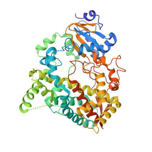
4K9V

Complex of CYP3A4 with a desoxyritonavir analog


Experimental Data Snapshot

  • Method: X-RAY DIFFRACTION
  • Resolution: 2.60 Å
  • R-Value Free: 0.294 
  • R-Value Work: 0.213 
  • R-Value Observed: 0.217 

Starting Model: experimental
View more details

wwPDB Validation   3D Report Full Report


Ligand Structure Quality Assessment 


This is version 1.5 of the entry. See complete history


Literature

Dissecting Cytochrome P450 3A4-Ligand Interactions Using Ritonavir Analogues.

Sevrioukova, I.F.Poulos, T.L.

(2013) Biochemistry 52: 4474-4481

  • DOI: https://doi.org/10.1021/bi4005396
  • Primary Citation of Related Structures:  
    4K9T, 4K9U, 4K9V, 4K9W, 4K9X

  • PubMed Abstract: 

    Cytochrome P450 3A4 (CYP3A4) inhibitors ritonavir and cobicistat, currently administered to HIV patients as pharmacoenhancers, were designed on the basis of the chemical structure/activity relationships rather than the CYP3A4 crystal structure. To better understand the structural basis for CYP3A4 inhibition and the ligand binding process, we investigated five desoxyritonavir analogues to elucidate how substitution/elimination of the phenyl side groups (Phe-1 and Phe-2) and removal of the isopropyl-thiazole (IPT) moiety affect affinity, inhibitory potency, and the ligand binding mode. Our experimental and structural data indicate that the side group size reduction not only drastically lowers affinity and inhibitory potency for CYP3A4 but also leads to multiple binding modes. Regardless of the side group chemical nature and the number of molecules bound, the space adjacent to the 369-371 peptide and Arg105 (Phe-2 site) is always occupied and, hence, must be a critically important binding site. When possible, the ligands also try to fill the pocket near the I-helix (Phe-1 site), even if it causes steric hindrance. Extensive hydrophobic interactions established at the Phe-1 site improve inhibitory potency, whereas contacts provided by IPT might strengthen the Fe-N bond. Overall, however, the end group contributes less to the ligand association process, which, in contrast, is greatly facilitated by the polar interactions mediated by the active site Ser119.


  • Organizational Affiliation

    Departments of Molecular Biology and Biochemistry, University of California, Irvine, California 92697, USA. [email protected]


Macromolecules
Find similar proteins by:  (by identity cutoff)  |  3D Structure
Entity ID: 1
MoleculeChains Sequence LengthOrganismDetailsImage
Cytochrome P450 3A4487Homo sapiensMutation(s): 0 
Gene Names: CYP3A3CYP3A4
EC: 1.14.13 (PDB Primary Data), 1.14.13.157 (PDB Primary Data), 1.14.13.32 (PDB Primary Data), 1.14.13.67 (PDB Primary Data), 1.14.13.97 (PDB Primary Data), 1.14.14.73 (UniProt), 1.14.14.1 (UniProt), 1.14.14.56 (UniProt), 1.14.14.55 (UniProt)
UniProt & NIH Common Fund Data Resources
Find proteins for P08684 (Homo sapiens)
Explore P08684 
Go to UniProtKB:  P08684
PHAROS:  P08684
GTEx:  ENSG00000160868 
Entity Groups  
Sequence Clusters30% Identity50% Identity70% Identity90% Identity95% Identity100% Identity
UniProt GroupP08684
Sequence Annotations
Expand
  • Reference Sequence
Small Molecules
Ligands 2 Unique
IDChains Name / Formula / InChI Key2D Diagram3D Interactions
HEM
Query on HEM

Download Ideal Coordinates CCD File 
B [auth A]PROTOPORPHYRIN IX CONTAINING FE
C34 H32 Fe N4 O4
KABFMIBPWCXCRK-RGGAHWMASA-L
6AW
Query on 6AW

Download Ideal Coordinates CCD File 
C [auth A]1,3-thiazol-5-ylmethyl [(3S,6S)-6-{[N-(methyl{[2-(propan-2-yl)-1,3-thiazol-4-yl]methyl}carbamoyl)-L-seryl]amino}octan-3-yl]carbamate
C25 H40 N6 O5 S2
SUQJOWRIFVHNKN-WFXMLNOXSA-N
Binding Affinity Annotations 
IDSourceBinding Affinity
6AW BindingDB:  4K9V Kd: 2300 (nM) from 1 assay(s)
IC50: 9700 (nM) from 1 assay(s)
Experimental Data & Validation

Experimental Data

  • Method: X-RAY DIFFRACTION
  • Resolution: 2.60 Å
  • R-Value Free: 0.294 
  • R-Value Work: 0.213 
  • R-Value Observed: 0.217 
  • Space Group: I 2 2 2
Unit Cell:
Length ( Å )Angle ( ˚ )
a = 77.62α = 90
b = 100.55β = 90
c = 130.04γ = 90
Software Package:
Software NamePurpose
Blu-Icedata collection
PHASERphasing
REFMACrefinement
MOSFLMdata reduction
SCALAdata scaling

Structure Validation

View Full Validation Report


Ligand Structure Quality Assessment 


Entry History 

Deposition Data

Revision History  (Full details and data files)

  • Version 1.0: 2013-06-19
    Type: Initial release
  • Version 1.1: 2013-06-26
    Changes: Database references
  • Version 1.2: 2013-07-17
    Changes: Database references
  • Version 1.3: 2014-10-29
    Changes: Structure summary
  • Version 1.4: 2017-10-25
    Changes: Author supporting evidence
  • Version 1.5: 2023-09-20
    Changes: Data collection, Database references, Derived calculations, Refinement description