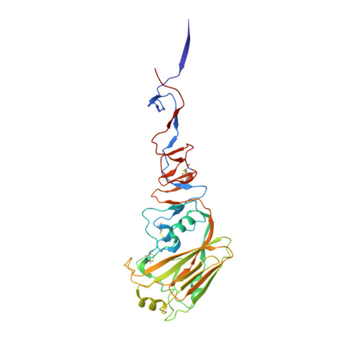
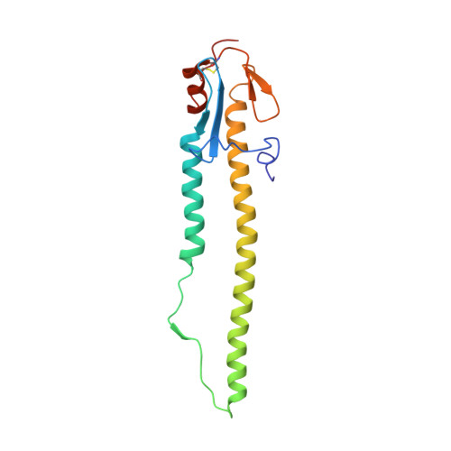
4JU0

Crystal structure of 2009 pandemic influenza virus hemagglutinin mutant D225E complexed with human receptor analogue LSTc


Experimental Data Snapshot

  • Method: X-RAY DIFFRACTION
  • Resolution: 2.91 Å
  • R-Value Free: 0.283 
  • R-Value Work: 0.240 
  • R-Value Observed: 0.243 

Starting Model: experimental
View more details

wwPDB Validation   3D Report Full Report


Ligand Structure Quality Assessment 


This is version 2.2 of the entry. See complete history


Literature

Molecular basis of the receptor binding specificity switch of the hemagglutinins from both the 1918 and 2009 pandemic influenza A viruses by a D225G substitution

Zhang, W.Shi, Y.Qi, J.Gao, F.Li, Q.Fan, Z.Yan, J.Gao, G.F.

(2013) J Virol 87: 5949-5958

  • DOI: https://doi.org/10.1128/JVI.00545-13
  • Primary Citation of Related Structures:  
    4JTV, 4JTX, 4JU0, 4JUG, 4JUH, 4JUJ

  • PubMed Abstract: 

    Influenza A virus uses sialic acids as cell entry receptors, and there are two main receptor forms, α2,6 linkage or α2,3 linkage to galactose, that determine virus host ranges (mammalian or avian). The receptor binding hemagglutinins (HAs) of both 1918 and 2009 pandemic H1N1 (18H1 and 09H1, respectively) influenza A viruses preferentially bind to the human α2,6 linkage receptor. A single D225G mutation in both H1s switches receptor binding specificity from α2,6 linkage binding to dual receptor binding. However, the molecular basis for this specificity switch is not fully understood. Here, we show via H1-ligand complex structures that the D225G substitution results in a loss of a salt bridge between amino acids D225 and K222, enabling the key residue Q226 to interact with the avian receptor, thereby obtaining dual receptor binding. This is further confirmed by a D225E mutant that retains human receptor binding specificity with the salt bridge intact.


  • Organizational Affiliation

    CAS Key Laboratory of Pathogenic Microbiology and Immunology, Institute of Microbiology, Chinese Academy of Sciences, Beijing, China.


Macromolecules
Find similar proteins by:  (by identity cutoff)  |  3D Structure
Entity ID: 1
MoleculeChains Sequence LengthOrganismDetailsImage
Hemagglutinin
A, C, E, G, I
A, C, E, G, I, K
322Influenza A virus (A/California/04/2009(H1N1))Mutation(s): 1 
Gene Names: HA
UniProt
Find proteins for C3W5S1 (Influenza A virus (strain swl A/California/04/2009 H1N1))
Explore C3W5S1 
Go to UniProtKB:  C3W5S1
Entity Groups  
Sequence Clusters30% Identity50% Identity70% Identity90% Identity95% Identity100% Identity
UniProt GroupC3W5S1
Glycosylation
Glycosylation Sites: 1
Sequence Annotations
Expand
  • Reference Sequence
Find similar proteins by:  (by identity cutoff)  |  3D Structure
Entity ID: 2
MoleculeChains Sequence LengthOrganismDetailsImage
Hemagglutinin
B, D, F, H, J
B, D, F, H, J, L
164Influenza A virus (A/California/04/2009(H1N1))Mutation(s): 0 
Gene Names: HA
UniProt
Find proteins for C3W5S1 (Influenza A virus (strain swl A/California/04/2009 H1N1))
Explore C3W5S1 
Go to UniProtKB:  C3W5S1
Entity Groups  
Sequence Clusters30% Identity50% Identity70% Identity90% Identity95% Identity100% Identity
UniProt GroupC3W5S1
Sequence Annotations
Expand
  • Reference Sequence
Oligosaccharides

Help

Entity ID: 3
MoleculeChains Length2D Diagram Glycosylation3D Interactions
N-acetyl-alpha-neuraminic acid-(2-6)-beta-D-galactopyranose-(1-4)-2-acetamido-2-deoxy-beta-D-glucopyranose-(1-3)-beta-D-galactopyranose
M, N, P, Q, R
4N/A
Glycosylation Resources
GlyTouCan:  G03509EL
GlyCosmos:  G03509EL
GlyGen:  G03509EL
Entity ID: 4
MoleculeChains Length2D Diagram Glycosylation3D Interactions
2-acetamido-2-deoxy-beta-D-glucopyranose-(1-4)-2-acetamido-2-deoxy-beta-D-glucopyranose
O
2N-Glycosylation
Glycosylation Resources
GlyTouCan:  G42666HT
GlyCosmos:  G42666HT
GlyGen:  G42666HT
Small Molecules
Ligands 2 Unique
IDChains Name / Formula / InChI Key2D Diagram3D Interactions
SIA
Query on SIA

Download Ideal Coordinates CCD File 
Y [auth K]N-acetyl-alpha-neuraminic acid
C11 H19 N O9
SQVRNKJHWKZAKO-YRMXFSIDSA-N
NAG
Query on NAG

Download Ideal Coordinates CCD File 
S [auth C]
T [auth C]
U [auth G]
V [auth I]
W [auth K]
S [auth C],
T [auth C],
U [auth G],
V [auth I],
W [auth K],
X [auth K]
2-acetamido-2-deoxy-beta-D-glucopyranose
C8 H15 N O6
OVRNDRQMDRJTHS-FMDGEEDCSA-N
Experimental Data & Validation

Experimental Data

  • Method: X-RAY DIFFRACTION
  • Resolution: 2.91 Å
  • R-Value Free: 0.283 
  • R-Value Work: 0.240 
  • R-Value Observed: 0.243 
  • Space Group: P 1
Unit Cell:
Length ( Å )Angle ( ˚ )
a = 66.871α = 62.06
b = 116.809β = 77.76
c = 116.503γ = 81.54
Software Package:
Software NamePurpose
MAR345dtbdata collection
PHASERphasing
PHENIXrefinement
HKL-2000data reduction
HKL-2000data scaling

Structure Validation

View Full Validation Report


Ligand Structure Quality Assessment 


Entry History 

Deposition Data

Revision History  (Full details and data files)

  • Version 1.0: 2013-05-01
    Type: Initial release
  • Version 1.1: 2013-08-07
    Changes: Database references
  • Version 2.0: 2020-07-29
    Type: Remediation
    Reason: Carbohydrate remediation
    Changes: Advisory, Atomic model, Data collection, Database references, Derived calculations, Structure summary
  • Version 2.1: 2023-11-08
    Changes: Data collection, Database references, Refinement description, Structure summary
  • Version 2.2: 2024-11-13
    Changes: Structure summary