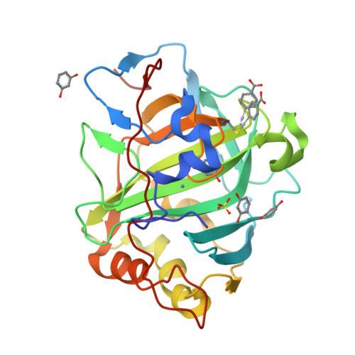
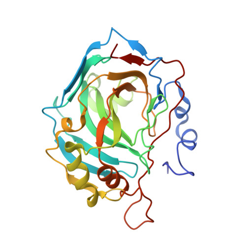
RCSB PDB - 4E49: Nucleophile recognition as an alternative inhibition mode for benzoic acid based carbonic anhydrase inhibitors

 4E49

Nucleophile recognition as an alternative inhibition mode for benzoic acid based carbonic anhydrase inhibitors


Experimental Data Snapshot

  • Method: X-RAY DIFFRACTION
  • Resolution: 1.45 Å
  • R-Value Free: 0.185 
  • R-Value Work: 0.158 
  • R-Value Observed: 0.160 

Starting Model: experimental
View more details

wwPDB Validation   3D Report Full Report


Ligand Structure Quality Assessment 

Created with Raphaël 2.3.0Worse 01 BetterLigand structure goodness of fit to experimental dataBest fitted MBOClick on this verticalbar to view details

This is version 1.1 of the entry. See complete history


Literature

Nucleophile recognition as an alternative inhibition mode for benzoic acid based carbonic anhydrase inhibitors

Martin, D.P.Cohen, S.M.

(2012) Chem Commun (Camb) 48: 5259-5261

  • DOI: https://doi.org/10.1039/c2cc32013d
  • Primary Citation of Related Structures:  
    4E3D, 4E3F, 4E3G, 4E3H, 4E49, 4E4A

  • PubMed Abstract: 

    A series of hydroxybenzoic acid derivatives have shown inhibitory activity against carbonic anhydrase (CA). X-ray crystallography shows that these molecules inhibit not by binding the active site metal ion but by strong hydrogen bonding to the metal-bound water nucleophile. The binding mode observed for these molecules is distinct when compared to other non-metal-binding CA inhibitors.


  • Organizational Affiliation

    Department of Chemistry and Biochemistry, University of California, San Diego, 9500 Gilman Drive, La Jolla, California 92093-0358, USA.


Macromolecules
Find similar proteins by:  (by identity cutoff)  |  3D Structure
Entity ID: 1
MoleculeChains Sequence LengthOrganismDetailsImage
Carbonic anhydrase 2260Homo sapiensMutation(s): 0 
Gene Names: CA2
EC: 4.2.1.1 (PDB Primary Data), 4.2.1.69 (UniProt)
UniProt & NIH Common Fund Data Resources
Find proteins for P00918 (Homo sapiens)
Explore P00918 
Go to UniProtKB:  P00918
PHAROS:  P00918
GTEx:  ENSG00000104267 
Entity Groups  
Sequence Clusters30% Identity50% Identity70% Identity90% Identity95% Identity100% Identity
UniProt GroupP00918
Sequence Annotations
Expand
  • Reference Sequence
Small Molecules
Ligands 4 Unique
IDChains Name / Formula / InChI Key2D Diagram3D Interactions
MBO
Query on MBO

Download Ideal Coordinates CCD File 
C [auth A]MERCURIBENZOIC ACID
C7 H5 Hg O2
FVFZSVRSDNUCGG-UHFFFAOYSA-N
RCO
Query on RCO

Download Ideal Coordinates CCD File 
D [auth A],
E [auth A],
F [auth A],
G [auth A]
RESORCINOL
C6 H6 O2
GHMLBKRAJCXXBS-UHFFFAOYSA-N
SO4
Query on SO4

Download Ideal Coordinates CCD File 
H [auth A]SULFATE ION
O4 S
QAOWNCQODCNURD-UHFFFAOYSA-L
ZN
Query on ZN

Download Ideal Coordinates CCD File 
B [auth A]ZINC ION
Zn
PTFCDOFLOPIGGS-UHFFFAOYSA-N
Binding Affinity Annotations 
IDSourceBinding Affinity
SO4 BindingDB:  4E49 Ki: min: 8.90e+7, max: 3.00e+8 (nM) from 4 assay(s)
RCO BindingDB:  4E49 Ki: min: 7700, max: 9400 (nM) from 2 assay(s)
Experimental Data & Validation

Experimental Data

  • Method: X-RAY DIFFRACTION
  • Resolution: 1.45 Å
  • R-Value Free: 0.185 
  • R-Value Work: 0.158 
  • R-Value Observed: 0.160 
  • Space Group: P 1 21 1
Unit Cell:
Length ( Å )Angle ( ˚ )
a = 42.179α = 90
b = 41.502β = 104.02
c = 71.824γ = 90
Software Package:
Software NamePurpose
APEXdata collection
PHASERphasing
REFMACrefinement
APEXdata reduction

Structure Validation

View Full Validation Report


Ligand Structure Quality Assessment 

Created with Raphaël 2.3.0Worse 01 BetterLigand structure goodness of fit to experimental dataBest fitted MBOClick on this verticalbar to view details

Entry History 

Deposition Data

Revision History  (Full details and data files)

  • Version 1.0: 2012-06-27
    Type: Initial release
  • Version 1.1: 2023-11-08
    Changes: Data collection, Database references, Derived calculations, Refinement description