4C1M

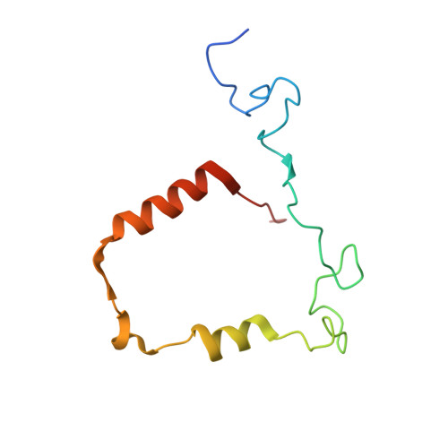
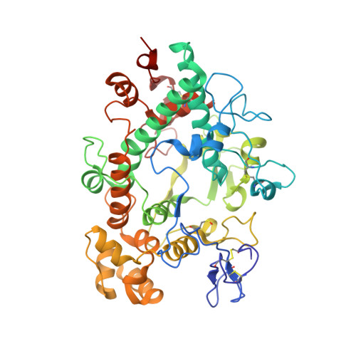
Myeloperoxidase in complex with the revesible inhibitor HX1


Experimental Data Snapshot

  • Method: X-RAY DIFFRACTION
  • Resolution: 2.00 Å
  • R-Value Free: 0.278 
  • R-Value Work: 0.222 
  • R-Value Observed: 0.225 

Starting Model: experimental
View more details

wwPDB Validation   3D Report Full Report


Ligand Structure Quality Assessment 


This is version 2.2 of the entry. See complete history


Literature

Potent Reversible Inhibition of Myeloperoxidase by Aromatic Hydroxamates

Forbes, L.V.Sjogren, T.Auchere, F.Jenkins, D.W.Thong, B.Laughton, D.Hemsley, P.Pairaudeau, G.Turner, R.Eriksson, H.Unitt, J.F.Kettle, A.J.

(2013) J Biol Chem 288: 36636

  • DOI: https://doi.org/10.1074/jbc.M113.507756
  • Primary Citation of Related Structures:  
    4C1M

  • PubMed Abstract: 

    The neutrophil enzyme myeloperoxidase (MPO) promotes oxidative stress in numerous inflammatory pathologies by producing hypohalous acids. Its inadvertent activity is a prime target for pharmacological control. Previously, salicylhydroxamic acid was reported to be a weak reversible inhibitor of MPO. We aimed to identify related hydroxamates that are good inhibitors of the enzyme. We report on three hydroxamates as the first potent reversible inhibitors of MPO. The chlorination activity of purified MPO was inhibited by 50% by a 5 nm concentration of a trifluoromethyl-substituted aromatic hydroxamate, HX1. The hydroxamates were specific for MPO in neutrophils and more potent toward MPO compared with a broad range of redox enzymes and alternative targets. Surface plasmon resonance measurements showed that the strength of binding of hydroxamates to MPO correlated with the degree of enzyme inhibition. The crystal structure of MPO-HX1 revealed that the inhibitor was bound within the active site cavity above the heme and blocked the substrate channel. HX1 was a mixed-type inhibitor of the halogenation activity of MPO with respect to both hydrogen peroxide and halide. Spectral analyses demonstrated that hydroxamates can act variably as substrates for MPO and convert the enzyme to a nitrosyl ferrous intermediate. This property was unrelated to their ability to inhibit MPO. We propose that aromatic hydroxamates bind tightly to the active site of MPO and prevent it from producing hypohalous acids. This mode of reversible inhibition has potential for blocking the activity of MPO and limiting oxidative stress during inflammation.


  • Organizational Affiliation

    From the Centre for Free Radical Research, Department of Pathology, University of Otago Christchurch, Christchurch 8140, New Zealand.


Macromolecules
Find similar proteins by:  (by identity cutoff)  |  3D Structure
Entity ID: 1
MoleculeChains Sequence LengthOrganismDetailsImage
MYELOPEROXIDASE LIGHT CHAIN
A, B
108Homo sapiensMutation(s): 0 
EC: 1.11.2.2 (PDB Primary Data), 1.11.1.7 (PDB Primary Data)
UniProt & NIH Common Fund Data Resources
Find proteins for P05164 (Homo sapiens)
Explore P05164 
Go to UniProtKB:  P05164
PHAROS:  P05164
GTEx:  ENSG00000005381 
Entity Groups  
Sequence Clusters30% Identity50% Identity70% Identity90% Identity95% Identity100% Identity
UniProt GroupP05164
Sequence Annotations
Expand
  • Reference Sequence
Find similar proteins by:  (by identity cutoff)  |  3D Structure
Entity ID: 2
MoleculeChains Sequence LengthOrganismDetailsImage
MYELOPEROXIDASE HEAVY CHAIN
C, D
467Homo sapiensMutation(s): 0 
EC: 1.11.2.2 (PDB Primary Data), 1.11.1.7 (PDB Primary Data)
UniProt & NIH Common Fund Data Resources
Find proteins for P05164 (Homo sapiens)
Explore P05164 
Go to UniProtKB:  P05164
PHAROS:  P05164
GTEx:  ENSG00000005381 
Entity Groups  
Sequence Clusters30% Identity50% Identity70% Identity90% Identity95% Identity100% Identity
UniProt GroupP05164
Glycosylation
Glycosylation Sites: 3Go to GlyGen: P05164-1
Sequence Annotations
Expand
  • Reference Sequence
Oligosaccharides

Help

Entity ID: 3
MoleculeChains Length2D Diagram Glycosylation3D Interactions
alpha-D-mannopyranose-(1-4)-[alpha-D-mannopyranose-(1-6)]beta-D-mannopyranose-(1-4)-2-acetamido-2-deoxy-beta-D-glucopyranose-(1-4)-[alpha-L-fucopyranose-(1-6)]2-acetamido-2-deoxy-beta-D-glucopyranose
E, F
6N-Glycosylation
Glycosylation Resources
GlyTouCan:  G70185OM
GlyCosmos:  G70185OM
GlyGen:  G70185OM
Small Molecules
Ligands 8 Unique
IDChains Name / Formula / InChI Key2D Diagram3D Interactions
HEM
Query on HEM

Download Ideal Coordinates CCD File 
G [auth A],
I [auth B]
PROTOPORPHYRIN IX CONTAINING FE
C34 H32 Fe N4 O4
KABFMIBPWCXCRK-RGGAHWMASA-L
NIH
Query on NIH

Download Ideal Coordinates CCD File 
H [auth A],
J [auth B]
2-{[3,5-BIS(TRIFLUOROMETHYL)BENZYL]AMINO}-N-HYDROXY-6-OXO-1,6-DIHYDROPYRIMIDINE-5-CARBOXAMIDE
C14 H10 F6 N4 O3
FNNKXGWDBVPDKY-UHFFFAOYSA-N
NAG
Query on NAG

Download Ideal Coordinates CCD File 
AA [auth D],
R [auth C],
S [auth C],
Z [auth D]
2-acetamido-2-deoxy-beta-D-glucopyranose
C8 H15 N O6
OVRNDRQMDRJTHS-FMDGEEDCSA-N
SO4
Query on SO4

Download Ideal Coordinates CCD File 
K [auth B],
O [auth C]
SULFATE ION
O4 S
QAOWNCQODCNURD-UHFFFAOYSA-L
GOL
Query on GOL

Download Ideal Coordinates CCD File 
L [auth C],
T [auth D]
GLYCEROL
C3 H8 O3
PEDCQBHIVMGVHV-UHFFFAOYSA-N
ACT
Query on ACT

Download Ideal Coordinates CCD File 
P [auth C],
Q [auth C],
W [auth D],
X [auth D],
Y [auth D]
ACETATE ION
C2 H3 O2
QTBSBXVTEAMEQO-UHFFFAOYSA-M
CA
Query on CA

Download Ideal Coordinates CCD File 
M [auth C],
U [auth D]
CALCIUM ION
Ca
BHPQYMZQTOCNFJ-UHFFFAOYSA-N
CL
Query on CL

Download Ideal Coordinates CCD File 
N [auth C],
V [auth D]
CHLORIDE ION
Cl
VEXZGXHMUGYJMC-UHFFFAOYSA-M
Modified Residues  1 Unique
IDChains TypeFormula2D DiagramParent
CSO
Query on CSO
C, D
L-PEPTIDE LINKINGC3 H7 N O3 SCYS
Binding Affinity Annotations 
IDSourceBinding Affinity
NIH BindingDB:  4C1M Kd: 15.8 (nM) from 1 assay(s)
IC50: 5.01 (nM) from 1 assay(s)
PDBBind:  4C1M Kd: 15.8 (nM) from 1 assay(s)
Experimental Data & Validation

Experimental Data

  • Method: X-RAY DIFFRACTION
  • Resolution: 2.00 Å
  • R-Value Free: 0.278 
  • R-Value Work: 0.222 
  • R-Value Observed: 0.225 
  • Space Group: P 1 21 1
Unit Cell:
Length ( Å )Angle ( ˚ )
a = 111.279α = 90
b = 63.44β = 97.36
c = 92.381γ = 90
Software Package:
Software NamePurpose
REFMACrefinement
MOSFLMdata reduction
SCALEPACKdata scaling
MOLREPphasing

Structure Validation

View Full Validation Report


Ligand Structure Quality Assessment 


Entry History 

Deposition Data

Revision History  (Full details and data files)

  • Version 1.0: 2013-11-13
    Type: Initial release
  • Version 1.1: 2013-11-20
    Changes: Database references
  • Version 1.2: 2014-01-15
    Changes: Database references
  • Version 2.0: 2020-07-29
    Type: Remediation
    Reason: Carbohydrate remediation
    Changes: Atomic model, Data collection, Derived calculations, Other, Structure summary
  • Version 2.1: 2023-12-20
    Changes: Data collection, Database references, Refinement description, Structure summary
  • Version 2.2: 2024-11-13
    Changes: Structure summary