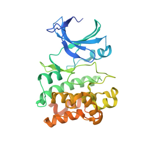
4BIB

Crystal Structures of Ask1-inhibitor Complexes


Experimental Data Snapshot

  • Method: X-RAY DIFFRACTION
  • Resolution: 2.43 Å
  • R-Value Free: 0.239 
  • R-Value Work: 0.201 
  • R-Value Observed: 0.203 

Starting Model: experimental
View more details

wwPDB Validation   3D Report Full Report


Ligand Structure Quality Assessment 


This is version 1.2 of the entry. See complete history


Literature

Crystal Structures of Ask1-Inhibtor Complexes Provide a Platform for Structure Based Drug Design.

Singh, O.Shillings, A.Craggs, P.Wall, I.Rowland, P.Skarzynski, T.Hobbs, C.I.Hardwick, P.Tanner, R.Blunt, M.Witty, D.R.Smith, K.J.

(2013) Protein Sci 22: 1071

  • DOI: https://doi.org/10.1002/pro.2298
  • Primary Citation of Related Structures:  
    4BF2, 4BHN, 4BIB, 4BIC, 4BID, 4BIE

  • PubMed Abstract: 

    ASK1, a member of the MAPK Kinase Kinase family of proteins has been shown to play a key role in cancer, neurodegeneration and cardiovascular diseases and is emerging as a possible drug target. Here we describe a 'replacement-soaking' method that has enabled the high-throughput X-ray structure determination of ASK1/ligand complexes. Comparison of the X-ray structures of five ASK1/ligand complexes from 3 different chemotypes illustrates that the ASK1 ATP binding site is able to accommodate a range of chemical diversity and different binding modes. The replacement-soaking system is also able to tolerate some protein flexibility. This crystal system provides a robust platform for ASK1/ligand structure determination and future structure based drug design.


  • Organizational Affiliation

    GlaxoSmithKline, Gunnels Wood Road, Stevenage, Hertfordshire, SG1 2NY, United Kingdom.


Macromolecules
Find similar proteins by:  (by identity cutoff)  |  3D Structure
Entity ID: 1
MoleculeChains Sequence LengthOrganismDetailsImage
MITOGEN-ACTIVATED PROTEIN KINASE KINASE KINASE 5
A, B
318Homo sapiensMutation(s): 1 
EC: 2.7.11.25
UniProt & NIH Common Fund Data Resources
Find proteins for Q99683 (Homo sapiens)
Explore Q99683 
Go to UniProtKB:  Q99683
PHAROS:  Q99683
GTEx:  ENSG00000197442 
Entity Groups  
Sequence Clusters30% Identity50% Identity70% Identity90% Identity95% Identity100% Identity
UniProt GroupQ99683
Sequence Annotations
Expand
  • Reference Sequence
Small Molecules
Ligands 3 Unique
IDChains Name / Formula / InChI Key2D Diagram3D Interactions
IEO
Query on IEO

Download Ideal Coordinates CCD File 
C [auth A],
H [auth B]
3-cyano-4-(piperidin-4-yloxy)-1H-indole-7-carboxamide
C15 H16 N4 O2
LJQBAOLMNHGSQC-UHFFFAOYSA-N
GOL
Query on GOL

Download Ideal Coordinates CCD File 
D [auth A],
F [auth A]
GLYCEROL
C3 H8 O3
PEDCQBHIVMGVHV-UHFFFAOYSA-N
ACT
Query on ACT

Download Ideal Coordinates CCD File 
E [auth A],
G [auth A],
I [auth B]
ACETATE ION
C2 H3 O2
QTBSBXVTEAMEQO-UHFFFAOYSA-M
Binding Affinity Annotations 
IDSourceBinding Affinity
IEO PDBBind:  4BIB IC50: 250 (nM) from 1 assay(s)
BindingDB:  4BIB IC50: 251 (nM) from 1 assay(s)
Experimental Data & Validation

Experimental Data

  • Method: X-RAY DIFFRACTION
  • Resolution: 2.43 Å
  • R-Value Free: 0.239 
  • R-Value Work: 0.201 
  • R-Value Observed: 0.203 
  • Space Group: P 65 2 2
Unit Cell:
Length ( Å )Angle ( ˚ )
a = 77.95α = 90
b = 77.95β = 90
c = 418.43γ = 120
Software Package:
Software NamePurpose
BUSTERrefinement
MOSFLMdata reduction
SCALAdata scaling
PHASERphasing

Structure Validation

View Full Validation Report


Ligand Structure Quality Assessment 


Entry History 

Deposition Data

Revision History  (Full details and data files)

  • Version 1.0: 2013-07-03
    Type: Initial release
  • Version 1.1: 2013-08-07
    Changes: Database references
  • Version 1.2: 2023-12-20
    Changes: Data collection, Database references, Derived calculations, Other, Refinement description