4B99 | pdb_00004b99

Crystal Structure of MAPK7 (ERK5) with inhibitor


Experimental Data Snapshot

  • Method: X-RAY DIFFRACTION
  • Resolution: 2.80 Å
  • R-Value Free: 
    0.287 (Depositor), 0.280 (DCC) 
  • R-Value Work: 
    0.222 (Depositor), 0.220 (DCC) 
  • R-Value Observed: 
    0.225 (Depositor) 

Starting Model: experimental
View more details

wwPDB Validation   3D Report Full Report


Ligand Structure Quality Assessment 

Created with Raphaël 2.3.0Worse 01 BetterLigand structure goodness of fit to experimental dataBest fitted R4LClick on this verticalbar to view details

This is version 1.4 of the entry. See complete history


Literature

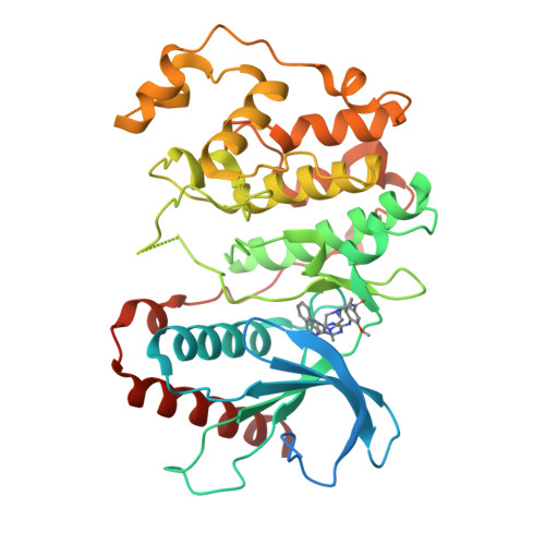
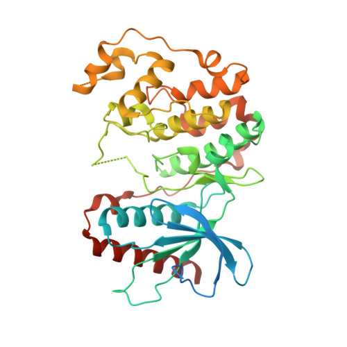
X-Ray Crystal Structure of Erk5 (Mapk7) in Complex with a Specific Inhibitor.

Elkins, J.M.Wang, J.Deng, X.Pattison, M.J.Arthur, S.Erazo, T.Gomez, N.Lizcano, J.M.Gray, N.S.Knapp, S.

(2013) J Med Chem 56: 4413

  • DOI: https://doi.org/10.1021/jm4000837
  • Primary Citation of Related Structures:  
    4B99

  • PubMed Abstract: 

    The protein kinase ERK5 (MAPK7) is an emerging drug target for a variety of indications, in particular for cancer where it plays a key role mediating cell proliferation, survival, epithelial-mesenchymal transition, and angiogenesis. To date, no three-dimensional structure has been published that would allow rational design of inhibitors. To address this, we determined the X-ray crystal structure of the human ERK5 kinase domain in complex with a highly specific benzo[e]pyrimido[5,4-b]diazepine-6(11H)-one inhibitor. The structure reveals that specific residue differences in the ATP-binding site, compared to the related ERKs p38s and JNKs, allow for the development of ERK5-specific inhibitors. The selectivity of previously observed ERK5 inhibitors can also be rationalized using this structure, which provides a template for future development of inhibitors with potential for treatment of disease.


  • Organizational Affiliation

    Structural Genomics Consortium, Nuffield Department of Clinical Medicine, University of Oxford, Old Road Campus Research Building, Roosevelt Drive, Oxford, OX3 7DQ, UK.


Macromolecules
Find similar proteins by:  (by identity cutoff)  |  3D Structure
Entity ID: 1
MoleculeChains Sequence LengthOrganismDetailsImage
MITOGEN-ACTIVATED PROTEIN KINASE 7398Homo sapiensMutation(s): 0 
EC: 2.7.11.24
UniProt & NIH Common Fund Data Resources
Find proteins for Q13164 (Homo sapiens)
Explore Q13164 
Go to UniProtKB:  Q13164
PHAROS:  Q13164
GTEx:  ENSG00000166484 
Entity Groups  
Sequence Clusters30% Identity50% Identity70% Identity90% Identity95% Identity100% Identity
UniProt GroupQ13164
Sequence Annotations
Expand
  • Reference Sequence
Small Molecules
Ligands 1 Unique
IDChains Name / Formula / InChI Key2D Diagram3D Interactions
R4L
Query on R4L

Download Ideal Coordinates CCD File 
B [auth A]11-cyclopentyl-2-[[2-methoxy-4-[4-(4-methylpiperazin-1-yl)piperidin-1-yl]carbonyl-phenyl]amino]-5-methyl-pyrimido[4,5-b][1,4]benzodiazepin-6-one
C35 H44 N8 O3
OXSKLFUMHBJZNS-UHFFFAOYSA-N
Binding Affinity Annotations 
IDSourceBinding Affinity
R4L BindingDB:  4B99 IC50: 162 (nM) from 1 assay(s)
EC50: 90 (nM) from 1 assay(s)
Experimental Data & Validation

Experimental Data

  • Method: X-RAY DIFFRACTION
  • Resolution: 2.80 Å
  • R-Value Free:  0.287 (Depositor), 0.280 (DCC) 
  • R-Value Work:  0.222 (Depositor), 0.220 (DCC) 
  • R-Value Observed: 0.225 (Depositor) 
Space Group: P 41 21 2
Unit Cell:
Length ( Å )Angle ( ˚ )
a = 95.15α = 90
b = 95.15β = 90
c = 119.45γ = 90
Software Package:
Software NamePurpose
REFMACrefinement
MOSFLMdata reduction
Aimlessdata scaling
PHASERphasing

Structure Validation

View Full Validation Report


Ligand Structure Quality Assessment 

Created with Raphaël 2.3.0Worse 01 BetterLigand structure goodness of fit to experimental dataBest fitted R4LClick on this verticalbar to view details

Entry History 

Revision History  (Full details and data files)

  • Version 1.0: 2012-09-19
    Type: Initial release
  • Version 1.1: 2013-05-22
    Changes: Database references
  • Version 1.2: 2013-06-26
    Changes: Database references
  • Version 1.3: 2018-01-24
    Changes: Structure summary
  • Version 1.4: 2023-12-20
    Changes: Data collection, Database references, Derived calculations, Other, Refinement description