4ACR | pdb_00004acr

Crystal structure of N-glycosylated, C-terminally truncated human glypican-1


Experimental Data Snapshot

  • Method: X-RAY DIFFRACTION
  • Resolution: 2.55 Å
  • R-Value Free: 
    0.292 (Depositor), 0.289 (DCC) 
  • R-Value Work: 
    0.251 (Depositor), 0.250 (DCC) 
  • R-Value Observed: 
    0.253 (Depositor) 

wwPDB Validation   3D Report Full Report


Ligand Structure Quality Assessment 

Created with Raphaël 2.3.0Worse 01 BetterLigand structure goodness of fit to experimental dataBest fitted NAGClick on this verticalbar to view details

This is version 1.4 of the entry. See complete history


Literature

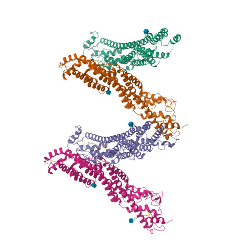
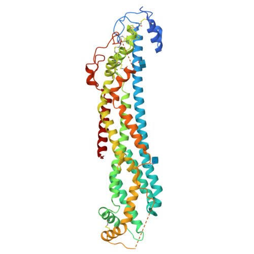
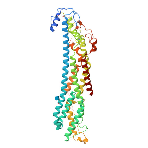
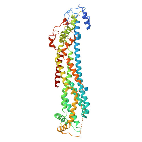
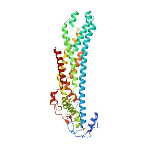
Crystal Structure of N-Glycosylated Human Glypican-1 Core Protein: Structure of Two Loops Evolutionarily Conserved in Vertebrate Glypican-1.

Svensson, G.Awad, W.Hakansson, M.Mani, K.Logan, D.T.

(2012) J Biological Chem 287: 14040

  • DOI: https://doi.org/10.1074/jbc.M111.322487
  • Primary Citation of Related Structures:  
    4ACR, 4AD7

  • PubMed Abstract: 

    Glypicans are a family of cell-surface proteoglycans that regulate Wnt, hedgehog, bone morphogenetic protein, and fibroblast growth factor signaling. Loss-of-function mutations in glypican core proteins and in glycosaminoglycan-synthesizing enzymes have revealed that glypican core proteins and their glycosaminoglycan chains are important in shaping animal development. Glypican core proteins consist of a stable α-helical domain containing 14 conserved Cys residues followed by a glycosaminoglycan attachment domain that becomes exclusively substituted with heparan sulfate (HS) and presumably adopts a random coil conformation. Removal of the α-helical domain results in almost exclusive addition of the glycosaminoglycan chondroitin sulfate, suggesting that factors in the α-helical domain promote assembly of HS. Glypican-1 is involved in brain development and is one of six members of the vertebrate family of glypicans. We expressed and crystallized N-glycosylated human glypican-1 lacking HS and N-glycosylated glypican-1 lacking the HS attachment domain. The crystal structure of glypican-1 was solved using crystals of selenomethionine-labeled glypican-1 core protein lacking the HS domain. No additional electron density was observed for crystals of glypican-1 containing the HS attachment domain, and CD spectra of the two protein species were highly similar. The crystal structure of N-glycosylated human glypican-1 core protein at 2.5 Å, the first crystal structure of a vertebrate glypican, reveals the complete disulfide bond arrangement of the conserved Cys residues, and it also extends the structural knowledge of glypicans for one α-helix and two long loops. Importantly, the loops are evolutionarily conserved in vertebrate glypican-1, and one of them is involved in glycosaminoglycan class determination.


  • Organizational Affiliation

    Department of Experimental Medical Science, Division of Neuroscience, Glycobiology Group, Lund University, Biomedical Center A13, SE-221 84 Lund, Sweden.


Macromolecules
Find similar proteins by:  (by identity cutoff)  |  3D Structure
Entity ID: 1
MoleculeChains Sequence LengthOrganismDetailsImage
GLYPICAN-1
A, B, C, D
478Homo sapiensMutation(s): 0 
UniProt & NIH Common Fund Data Resources
Find proteins for P35052 (Homo sapiens)
Explore P35052 
Go to UniProtKB:  P35052
PHAROS:  P35052
GTEx:  ENSG00000063660 
Entity Groups  
Sequence Clusters30% Identity50% Identity70% Identity90% Identity95% Identity100% Identity
UniProt GroupP35052
Glycosylation
Glycosylation Sites: 1Go to GlyGen: P35052-1
Sequence Annotations
Expand
  • Reference Sequence
Experimental Data & Validation

Experimental Data

  • Method: X-RAY DIFFRACTION
  • Resolution: 2.55 Å
  • R-Value Free:  0.292 (Depositor), 0.289 (DCC) 
  • R-Value Work:  0.251 (Depositor), 0.250 (DCC) 
  • R-Value Observed: 0.253 (Depositor) 
Space Group: P 1 21 1
Unit Cell:
Length ( Å )Angle ( ˚ )
a = 47.17α = 90
b = 168.63β = 94.59
c = 147.76γ = 90
Software Package:
Software NamePurpose
PHENIXrefinement
XDSdata reduction
XSCALEdata scaling
Auto-Rickshawphasing

Structure Validation

View Full Validation Report


Ligand Structure Quality Assessment 

Created with Raphaël 2.3.0Worse 01 BetterLigand structure goodness of fit to experimental dataBest fitted NAGClick on this verticalbar to view details

Entry History 

Deposition Data

Revision History  (Full details and data files)

  • Version 1.0: 2012-02-22
    Type: Initial release
  • Version 1.1: 2012-05-02
    Changes: Other
  • Version 1.2: 2018-01-17
    Changes: Data collection
  • Version 1.3: 2020-07-29
    Type: Remediation
    Reason: Carbohydrate remediation
    Changes: Advisory, Data collection, Derived calculations, Other, Structure summary
  • Version 1.4: 2024-11-20
    Changes: Data collection, Database references, Structure summary