3WMV

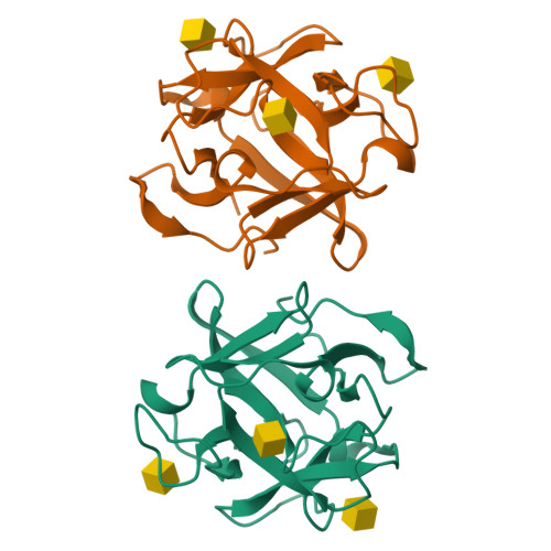
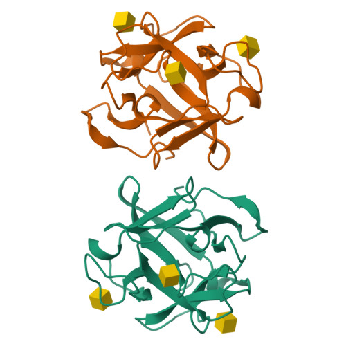
The structure of an anti-cancer lectin mytilec with ligand from the mussel Mytilus galloprovincialis


Experimental Data Snapshot

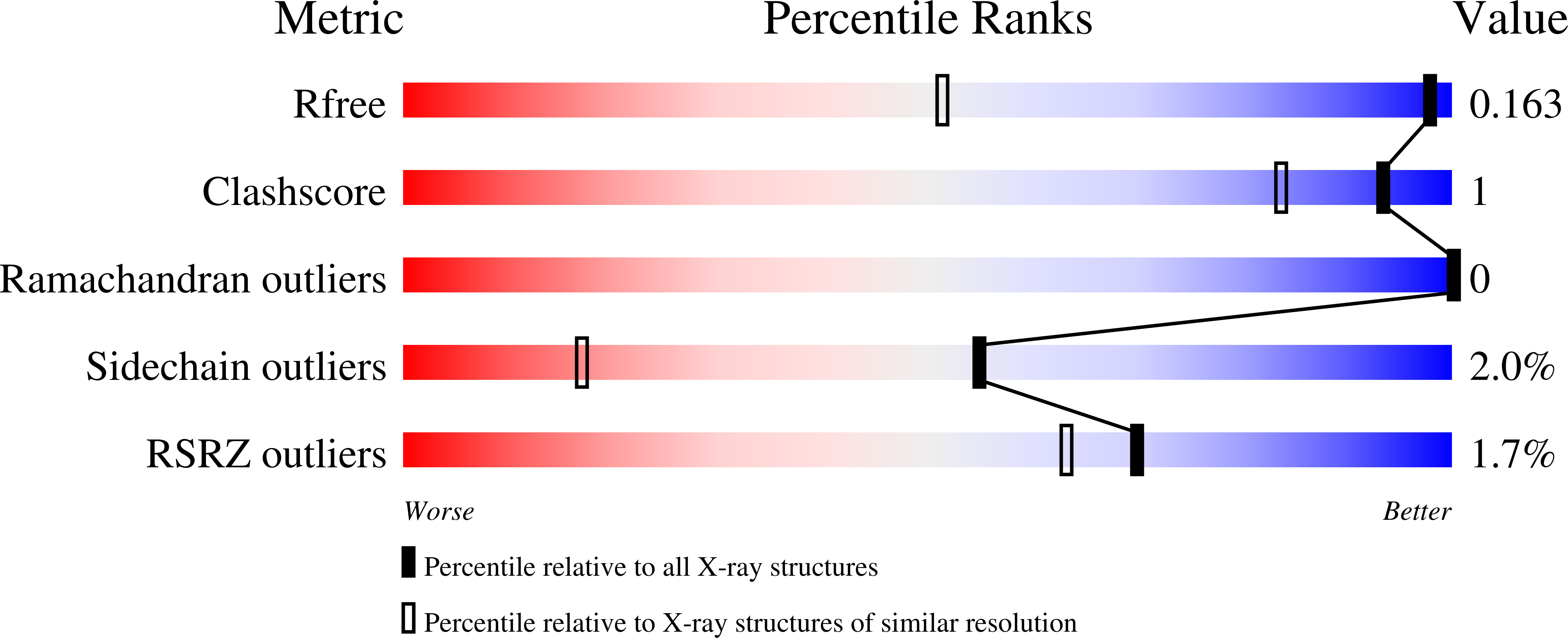
  • Method: X-RAY DIFFRACTION
  • Resolution: 1.05 Å
  • R-Value Free: 0.155 
  • R-Value Work: 0.136 
  • R-Value Observed: 0.137 

wwPDB Validation   3D Report Full Report


Ligand Structure Quality Assessment 


This is version 2.1 of the entry. See complete history


Literature

Crystal structure of MytiLec, a galactose-binding lectin from the mussel Mytilus galloprovincialis with cytotoxicity against certain cancer cell types

Terada, D.Kawai, F.Noguchi, H.Unzai, S.Hasan, I.Fujii, Y.Park, S.-Y.Ozeki, Y.Tame, J.R.H.

(2016) Sci Rep 6: 28344-28344

  • DOI: https://doi.org/10.1038/srep28344
  • Primary Citation of Related Structures:  
    3WMU, 3WMV

  • PubMed Abstract: 

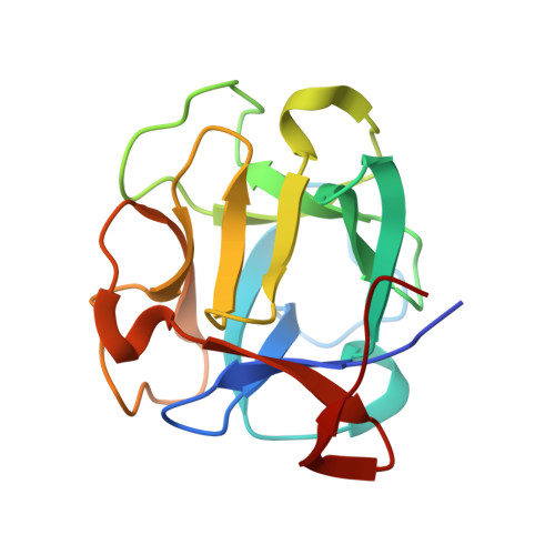
    MytiLec is a lectin, isolated from bivalves, with cytotoxic activity against cancer cell lines that express globotriaosyl ceramide, Galα(1,4)Galβ(1,4)Glcα1-Cer, on the cell surface. Functional analysis shows that the protein binds to the disaccharide melibiose, Galα(1,6)Glc, and the trisaccharide globotriose, Galα(1,4)Galβ(1,4)Glc. Recombinant MytiLec expressed in bacteria showed the same haemagglutinating and cytotoxic activity against Burkitt's lymphoma (Raji) cells as the native form. The crystal structure has been determined to atomic resolution, in the presence and absence of ligands, showing the protein to be a member of the β-trefoil family, but with a mode of ligand binding unique to a small group of related trefoil lectins. Each of the three pseudo-equivalent binding sites within the monomer shows ligand binding, and the protein forms a tight dimer in solution. An engineered monomer mutant lost all cytotoxic activity against Raji cells, but retained some haemagglutination activity, showing that the quaternary structure of the protein is important for its cellular effects.


  • Organizational Affiliation

    Graduate School of Medical Life Science, Yokohama City University, 1-7-29 Suehiro, Yokohama, Kanagawa 230-0045, Japan.


Macromolecules
Find similar proteins by:  (by identity cutoff)  |  3D Structure
Entity ID: 1
MoleculeChains Sequence LengthOrganismDetailsImage
Lectin
A, B
153Mytilus galloprovincialisMutation(s): 1 
UniProt
Find proteins for B3EWR1 (Mytilus galloprovincialis)
Explore B3EWR1 
Go to UniProtKB:  B3EWR1
Entity Groups  
Sequence Clusters30% Identity50% Identity70% Identity90% Identity95% Identity100% Identity
UniProt GroupB3EWR1
Sequence Annotations
Expand
  • Reference Sequence
Experimental Data & Validation

Experimental Data

  • Method: X-RAY DIFFRACTION
  • Resolution: 1.05 Å
  • R-Value Free: 0.155 
  • R-Value Work: 0.136 
  • R-Value Observed: 0.137 
  • Space Group: C 1 2 1
Unit Cell:
Length ( Å )Angle ( ˚ )
a = 80.276α = 90
b = 64.215β = 121.51
c = 74.282γ = 90
Software Package:
Software NamePurpose
HKL-2000data collection
MOLREPphasing
REFMACrefinement
HKL-2000data reduction
HKL-2000data scaling

Structure Validation

View Full Validation Report


Ligand Structure Quality Assessment 


Entry History 

Deposition Data

Revision History  (Full details and data files)

  • Version 1.0: 2014-12-03
    Type: Initial release
  • Version 1.1: 2017-06-07
    Changes: Database references
  • Version 2.0: 2020-07-29
    Type: Remediation
    Reason: Carbohydrate remediation
    Changes: Atomic model, Data collection, Database references, Derived calculations, Structure summary
  • Version 2.1: 2024-03-20
    Changes: Data collection, Database references, Structure summary