3QND | pdb_00003qnd

crystal structure of Ad37 fiber knob in complex with trivalent sialic acid inhibitor


Experimental Data Snapshot

  • Method: X-RAY DIFFRACTION
  • Resolution: 2.40 Å
  • R-Value Free: 
    0.263 (Depositor), 0.260 (DCC) 
  • R-Value Work: 
    0.221 (Depositor), 0.220 (DCC) 
  • R-Value Observed: 
    0.223 (Depositor) 

Starting Model: experimental
View more details

wwPDB Validation   3D Report Full Report


Ligand Structure Quality Assessment 

Created with Raphaël 2.3.0Worse 01 BetterLigand structure goodness of fit to experimental dataBest fitted SIAClick on this verticalbar to view details

This is version 1.4 of the entry. See complete history


Literature

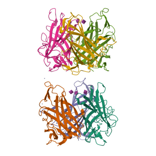
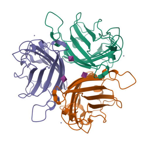
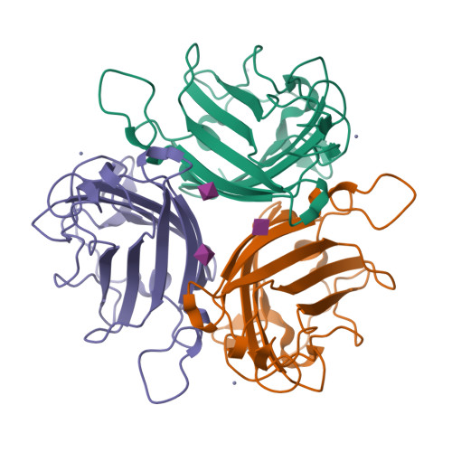
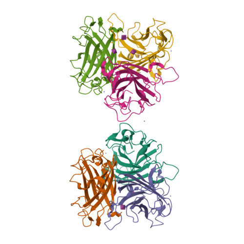
A potent trivalent sialic Acid inhibitor of adenovirus type 37 infection of human corneal cells.

Spjut, S.Qian, W.Bauer, J.Storm, R.Frangsmyr, L.Stehle, T.Arnberg, N.Elofsson, M.

(2011) Angew Chem Int Ed Engl 50: 6519-6521


Macromolecules
Find similar proteins by:  (by identity cutoff)  |  3D Structure
Entity ID: 1
MoleculeChains Sequence LengthOrganismDetailsImage
Fiber protein194Human adenovirus D37Mutation(s): 0 
Gene Names: L5
UniProt
Find proteins for Q64823 (Human adenovirus D37)
Explore Q64823 
Go to UniProtKB:  Q64823
Entity Groups  
Sequence Clusters30% Identity50% Identity70% Identity90% Identity95% Identity100% Identity
UniProt GroupQ64823
Sequence Annotations
Expand
  • Reference Sequence
Small Molecules
Ligands 2 Unique
IDChains Name / Formula / InChI Key2D Diagram3D Interactions
SIA
Query on SIA

Download Ideal Coordinates CCD File 
H [auth A]
J [auth B]
L [auth C]
O [auth E]
Q [auth F]
N-acetyl-alpha-neuraminic acid
C11 H19 N O9
SQVRNKJHWKZAKO-YRMXFSIDSA-N
ZN
Query on ZN

Download Ideal Coordinates CCD File 
G [auth A]
I [auth B]
K [auth C]
M [auth E]
N [auth E]
ZINC ION
Zn
PTFCDOFLOPIGGS-UHFFFAOYSA-N
Experimental Data & Validation

Experimental Data

  • Method: X-RAY DIFFRACTION
  • Resolution: 2.40 Å
  • R-Value Free:  0.263 (Depositor), 0.260 (DCC) 
  • R-Value Work:  0.221 (Depositor), 0.220 (DCC) 
  • R-Value Observed: 0.223 (Depositor) 
Space Group: P 21 21 21
Unit Cell:
Length ( Å )Angle ( ˚ )
a = 105.19α = 90
b = 108.645β = 90
c = 112.511γ = 90
Software Package:
Software NamePurpose
XDSdata scaling
PHASERphasing
REFMACrefinement
XDSdata reduction

Structure Validation

View Full Validation Report


Ligand Structure Quality Assessment 

Created with Raphaël 2.3.0Worse 01 BetterLigand structure goodness of fit to experimental dataBest fitted SIAClick on this verticalbar to view details

Entry History 

Deposition Data

Revision History  (Full details and data files)

  • Version 1.0: 2011-06-15
    Type: Initial release
  • Version 1.1: 2011-07-13
    Changes: Version format compliance
  • Version 1.2: 2011-08-10
    Changes: Database references
  • Version 1.3: 2020-07-29
    Type: Remediation
    Reason: Carbohydrate remediation
    Changes: Data collection, Database references, Derived calculations, Structure summary
  • Version 1.4: 2023-09-13
    Changes: Data collection, Database references, Refinement description, Structure summary