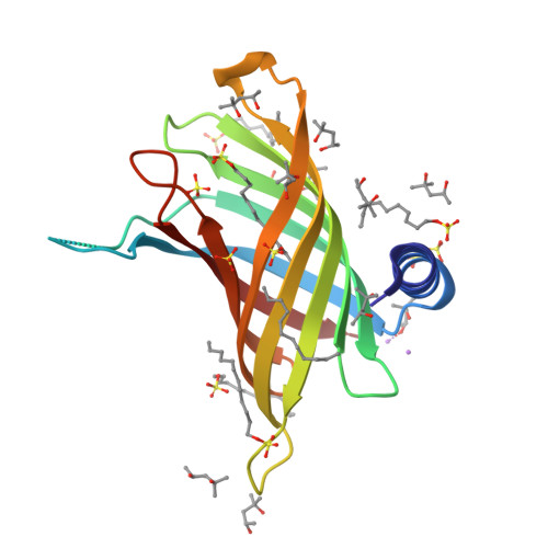
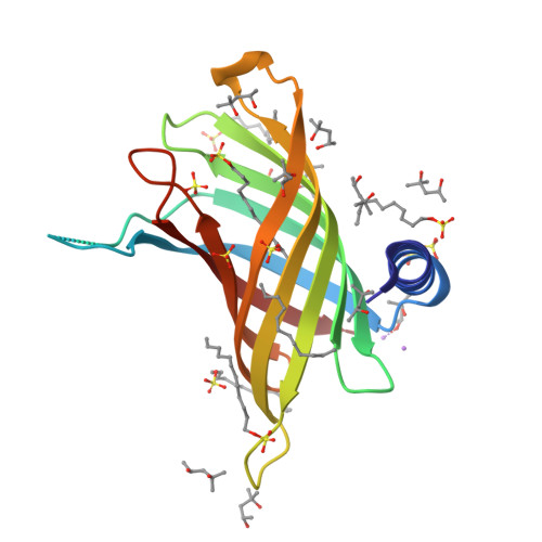
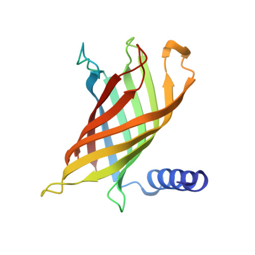
3GP6

Crystal structure of PagP in SDS/MPD


Experimental Data Snapshot

  • Method: X-RAY DIFFRACTION
  • Resolution: 1.40 Å
  • R-Value Free: 0.208 
  • R-Value Work: 0.171 
  • R-Value Observed: 0.173 

Starting Model: experimental
View more details

wwPDB Validation   3D Report Full Report


Ligand Structure Quality Assessment 


This is version 1.2 of the entry. See complete history


Literature

PagP crystallized from SDS/cosolvent reveals the route for phospholipid access to the hydrocarbon ruler.

Cuesta-Seijo, J.A.Neale, C.Khan, M.A.Moktar, J.Tran, C.D.Bishop, R.E.Pomes, R.Prive, G.G.

(2010) Structure 18: 1210-1219

  • DOI: https://doi.org/10.1016/j.str.2010.06.014
  • Primary Citation of Related Structures:  
    3GP6

  • PubMed Abstract: 

    Enzymatic reactions involving bilayer lipids occur in an environment with strict physical and topological constraints. The integral membrane enzyme PagP transfers a palmitoyl group from a phospholipid to lipid A in order to assist Escherichia coli in evading host immune defenses during infection. PagP measures the palmitoyl group with an internal hydrocarbon ruler that is formed in the interior of the eight-stranded antiparallel β barrel. The access and egress of the palmitoyl group is thought to take a lateral route from the bilayer phase to the barrel interior. Molecular dynamics, mutagenesis, and a 1.4 A crystal structure of PagP in an SDS / 2-methyl-2,4-pentanediol (MPD) cosolvent system reveal that phospholipid access occurs at the crenel present between strands F and G of PagP. In this way, the phospholipid head group can remain exposed to the cell exterior while the lipid acyl chain remains in a predominantly hydrophobic environment as it translocates to the protein interior.


  • Organizational Affiliation

    Division of Cancer Genomics and Proteomics, Ontario Cancer Institute and Campbell Family Cancer Research Institute, 101 College Street, Toronto, ON M5G 1L7, Canada.


Macromolecules
Find similar proteins by:  (by identity cutoff)  |  3D Structure
Entity ID: 1
MoleculeChains Sequence LengthOrganismDetailsImage
Protein pagP163Escherichia coli K-12Mutation(s): 0 
Gene Names: b0622crcAJW0617ybeG
EC: 2.3.1.251
Membrane Entity: Yes 
UniProt
Find proteins for P37001 (Escherichia coli (strain K12))
Explore P37001 
Go to UniProtKB:  P37001
Entity Groups  
Sequence Clusters30% Identity50% Identity70% Identity90% Identity95% Identity100% Identity
UniProt GroupP37001
Sequence Annotations
Expand
  • Reference Sequence
Small Molecules
Ligands 5 Unique
IDChains Name / Formula / InChI Key2D Diagram3D Interactions
SDS
Query on SDS

Download Ideal Coordinates CCD File 
B [auth A]
C [auth A]
D [auth A]
E [auth A]
F [auth A]
B [auth A],
C [auth A],
D [auth A],
E [auth A],
F [auth A],
G [auth A]
DODECYL SULFATE
C12 H26 O4 S
MOTZDAYCYVMXPC-UHFFFAOYSA-N
MRD
Query on MRD

Download Ideal Coordinates CCD File 
Q [auth A](4R)-2-METHYLPENTANE-2,4-DIOL
C6 H14 O2
SVTBMSDMJJWYQN-RXMQYKEDSA-N
MPD
Query on MPD

Download Ideal Coordinates CCD File 
H [auth A]
I [auth A]
J [auth A]
K [auth A]
L [auth A]
H [auth A],
I [auth A],
J [auth A],
K [auth A],
L [auth A],
M [auth A],
N [auth A],
O [auth A],
P [auth A]
(4S)-2-METHYL-2,4-PENTANEDIOL
C6 H14 O2
SVTBMSDMJJWYQN-YFKPBYRVSA-N
SO4
Query on SO4

Download Ideal Coordinates CCD File 
R [auth A]
S [auth A]
T [auth A]
U [auth A]
V [auth A]
R [auth A],
S [auth A],
T [auth A],
U [auth A],
V [auth A],
W [auth A]
SULFATE ION
O4 S
QAOWNCQODCNURD-UHFFFAOYSA-L
LI
Query on LI

Download Ideal Coordinates CCD File 
X [auth A],
Y [auth A]
LITHIUM ION
Li
HBBGRARXTFLTSG-UHFFFAOYSA-N
Experimental Data & Validation

Experimental Data

  • Method: X-RAY DIFFRACTION
  • Resolution: 1.40 Å
  • R-Value Free: 0.208 
  • R-Value Work: 0.171 
  • R-Value Observed: 0.173 
  • Space Group: P 62 2 2
Unit Cell:
Length ( Å )Angle ( ˚ )
a = 113.228α = 90
b = 113.228β = 90
c = 55.064γ = 120
Software Package:
Software NamePurpose
HKL-3000data collection
EPMRphasing
REFMACrefinement
HKL-3000data reduction
SCALEPACKdata scaling

Structure Validation

View Full Validation Report


Ligand Structure Quality Assessment 


Entry History 

Deposition Data

Revision History  (Full details and data files)

  • Version 1.0: 2010-06-23
    Type: Initial release
  • Version 1.1: 2011-07-13
    Changes: Version format compliance
  • Version 1.2: 2023-09-06
    Changes: Data collection, Database references, Derived calculations, Refinement description