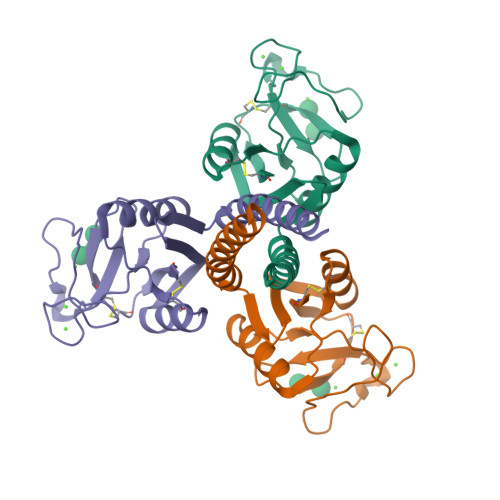
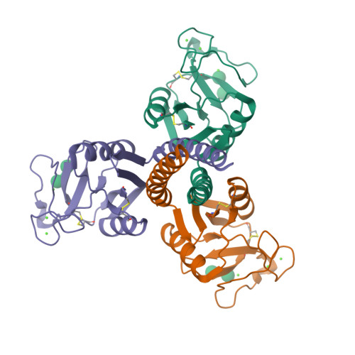
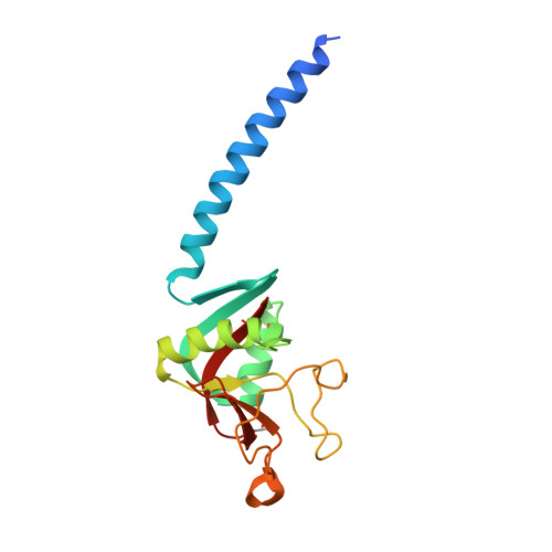
3G83

Crystal structure of the trimeric neck and carbohydrate recognition domain of human surfactant protein D in complex with alpha 1,2 dimannose.


Experimental Data Snapshot

  • Method: X-RAY DIFFRACTION
  • Resolution: 1.90 Å
  • R-Value Free: 0.215 
  • R-Value Work: 0.189 
  • R-Value Observed: 0.190 

Starting Model: experimental
View more details

wwPDB Validation   3D Report Full Report


This is version 2.2 of the entry. See complete history


Literature

Recognition of mannosylated ligands and influenza A virus by human surfactant protein D: contributions of an extended site and residue 343.

Crouch, E.Hartshorn, K.Horlacher, T.McDonald, B.Smith, K.Cafarella, T.Seaton, B.Seeberger, P.H.Head, J.

(2009) Biochemistry 48: 3335-3345

  • DOI: https://doi.org/10.1021/bi8022703
  • Primary Citation of Related Structures:  
    3G81, 3G83, 3G84

  • PubMed Abstract: 

    Surfactant protein D (SP-D) plays important roles in antiviral host defense. Although SP-D shows a preference for glucose/maltose, the protein also recognizes d-mannose and a variety of mannose-rich microbial ligands. This latter preference prompted an examination of the mechanisms of mannose recognition, particularly as they relate to high-mannose viral glycans. Trimeric neck plus carbohydrate recognition domains from human SP-D (hNCRD) preferred alpha1-2-linked dimannose (DM) over the branched trimannose (TM) core, alpha1-3 or alpha1-6 DM, or D-mannose. Previous studies have shown residues flanking the carbohydrate binding site can fine-tune ligand recognition. A mutant with valine at 343 (R343V) showed enhanced binding to mannan relative to wild type and R343A. No alteration in affinity was observed for D-mannose or for alpha1-3- or alpha1-6-linked DM; however, substantially increased affinity was observed for alpha1-2 DM. Both proteins showed efficient recognition of linear and branched subdomains of high-mannose glycans on carbohydrate microarrays, and R343V showed increased binding to a subset of the oligosaccharides. Crystallographic analysis of an R343V complex with 1,2-DM showed a novel mode of binding. The disaccharide is bound to calcium by the reducing sugar ring, and a stabilizing H-bond is formed between the 2-OH of the nonreducing sugar ring and Arg349. Although hNCRDs show negligible binding to influenza A virus (IAV), R343V showed markedly enhanced viral neutralizing activity. Hydrophobic substitutions for Arg343 selectively blocked binding of a monoclonal antibody (Hyb 246-05) that inhibits IAV binding activity. Our findings demonstrate an extended ligand binding site for mannosylated ligands and the significant contribution of the 343 side chain to specific recognition of multivalent microbial ligands, including high-mannose viral glycans.


  • Organizational Affiliation

    Department of Pathology and Immunology, Washington University School of Medicine, St. Louis, Missouri 63110, USA. [email protected]


Macromolecules
Find similar proteins by:  (by identity cutoff)  |  3D Structure
Entity ID: 1
MoleculeChains Sequence LengthOrganismDetailsImage
Pulmonary surfactant-associated protein D
A, B, C
160Homo sapiensMutation(s): 0 
Gene Names: SFTPDPSPDSFTP4
UniProt & NIH Common Fund Data Resources
Find proteins for P35247 (Homo sapiens)
Explore P35247 
Go to UniProtKB:  P35247
PHAROS:  P35247
GTEx:  ENSG00000133661 
Entity Groups  
Sequence Clusters30% Identity50% Identity70% Identity90% Identity95% Identity100% Identity
UniProt GroupP35247
Sequence Annotations
Expand
  • Reference Sequence
Oligosaccharides

Help

Entity ID: 2
MoleculeChains Length2D Diagram Glycosylation3D Interactions
alpha-D-mannopyranose-(1-2)-alpha-D-mannopyranose
D, E, F
2N/A
Glycosylation Resources
GlyTouCan:  G53402KW
GlyCosmos:  G53402KW
GlyGen:  G53402KW
Small Molecules
Ligands 1 Unique
IDChains Name / Formula / InChI Key2D Diagram3D Interactions
CA
Query on CA

Download Ideal Coordinates CCD File 
G [auth A]
H [auth A]
I [auth A]
J [auth B]
K [auth B]
G [auth A],
H [auth A],
I [auth A],
J [auth B],
K [auth B],
L [auth B],
M [auth C],
N [auth C],
O [auth C]
CALCIUM ION
Ca
BHPQYMZQTOCNFJ-UHFFFAOYSA-N
Biologically Interesting Molecules (External Reference) 1 Unique
Experimental Data & Validation

Experimental Data

  • Method: X-RAY DIFFRACTION
  • Resolution: 1.90 Å
  • R-Value Free: 0.215 
  • R-Value Work: 0.189 
  • R-Value Observed: 0.190 
  • Space Group: P 1 21 1
Unit Cell:
Length ( Å )Angle ( ˚ )
a = 55.754α = 90
b = 108.774β = 91.58
c = 56.011γ = 90
Software Package:
Software NamePurpose
DENZOdata reduction
SCALEPACKdata scaling
PHENIXrefinement
PDB_EXTRACTdata extraction
CBASSdata collection

Structure Validation

View Full Validation Report


Entry History 

Deposition Data

  • Released Date: 2009-03-17 
  • Deposition Author(s): Head, J.F.

Revision History  (Full details and data files)

  • Version 1.0: 2009-03-17
    Type: Initial release
  • Version 1.1: 2011-07-13
    Changes: Version format compliance
  • Version 1.2: 2017-11-01
    Changes: Refinement description
  • Version 2.0: 2020-07-29
    Type: Remediation
    Reason: Carbohydrate remediation
    Changes: Atomic model, Data collection, Database references, Derived calculations, Structure summary
  • Version 2.1: 2023-09-06
    Changes: Data collection, Database references, Refinement description, Structure summary
  • Version 2.2: 2024-11-06
    Changes: Structure summary