1TRP | pdb_00001trp

X-RAY CRYSTALLOGRAPHIC AND CALORIMERIC STUDIES OF THE EFFECTS OF THE MUTATION TRP 59 TYR IN RIBONUCLEASE T1


Experimental Data Snapshot

  • Method: X-RAY DIFFRACTION
  • Resolution: 2.40 Å
  • R-Value Work: 
    0.250 (DCC) 
  • R-Value Observed: 
    0.160 (Depositor) 

wwPDB Validation   3D Report Full Report


Ligand Structure Quality Assessment 

Created with Raphaël 2.3.0Worse 01 BetterLigand structure goodness of fit to experimental dataBest fitted 2GPClick on this verticalbar to view details

This is version 1.4 of the entry. See complete history


Literature

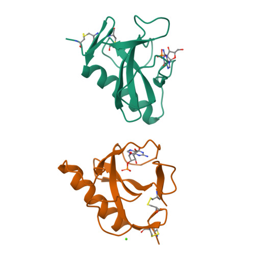
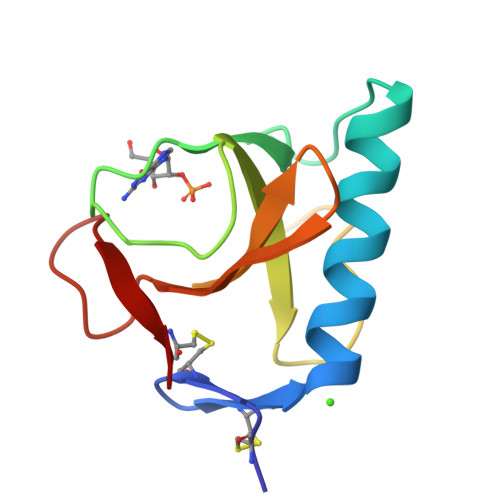
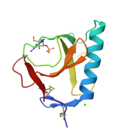
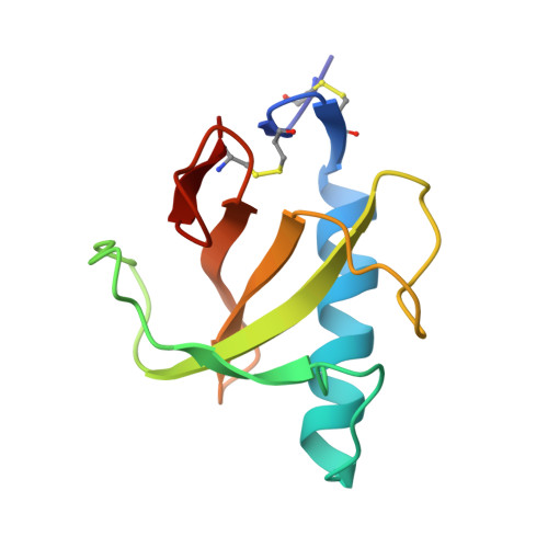
X-ray crystallographic and calorimetric studies of the effects of the mutation Trp59-->Tyr in ribonuclease T1.

Schubert, W.D.Schluckebier, G.Backmann, J.Granzin, J.Kisker, C.Choe, H.W.Hahn, U.Pfeil, W.Saenger, W.

(1994) Eur J Biochem 220: 527-534

  • DOI: https://doi.org/10.1111/j.1432-1033.1994.tb18652.x
  • Primary Citation of Related Structures:  
    1TRP, 1TRQ

  • PubMed Abstract: 

    Two mutants of ribonuclease T1 (RNaseT1), [59-tyrosine]ribonuclease T1 (W59Y) and [45-tryptophan,59-tyrosine]ribonuclease T1 (Y45W/W59Y) possess between 150% and 190% wild-type activity. They have been crystallised as complexes of the inhibitor 2'-guanylic acid and analysed by X-ray diffraction at resolutions of 0.23 nm and 0.24 nm, respectively. The space group for both is monoclinic, P2(1), with two molecules/asymmetric unit, W59Y: a = 4.934 nm, b = 4.820 nm, c = 4.025 nm, beta = 90.29 degrees. Y45W/W59Y: a = 4.915 nm, b = 4.815 nm, c = 4.015 nm, beta = 90.35 degrees. Compared to wild-type RNaseT1 in complex with 2'-guanylic acid (2'GMP) both mutant inhibitor complexes indicate that the replacement of Trp59 by Tyr leads to a 0.04-nm inward shift of the single alpha-helix and to significant differences in the active-site geometry, inhibitor conformation and inhibitor binding. Calorimetric studies of a range of mutants [24-tryptophan]ribonuclease T1 (Y24W), [42-tryptophan]ribonuclease T1 (Y42W), [45-tryptophan]ribonuclease T1 (Y45W), [92-alanine]ribonuclease T1 (H92A) and [92-threonine]ribonuclease T1 (H92T) with and without the further mutation Trp59-->Tyr showed that mutant proteins for which Trp59 is replaced by Tyr exhibit slightly decreased thermal stability.


  • Organizational Affiliation

    Institut für Kristallographie, Freien Universität Berlin, Germany.


Macromolecules
Find similar proteins by:  (by identity cutoff)  |  3D Structure
Entity ID: 1
MoleculeChains Sequence LengthOrganismDetailsImage
RIBONUCLEASE T1 ISOZYME
A, B
104Aspergillus oryzaeMutation(s): 0 
EC: 3.1.27.3 (PDB Primary Data), 4.6.1.24 (UniProt)
UniProt
Find proteins for P00651 (Aspergillus oryzae (strain ATCC 42149 / RIB 40))
Explore P00651 
Go to UniProtKB:  P00651
Entity Groups  
Sequence Clusters30% Identity50% Identity70% Identity90% Identity95% Identity100% Identity
UniProt GroupP00651
Sequence Annotations
Expand
  • Reference Sequence
Experimental Data & Validation

Experimental Data

  • Method: X-RAY DIFFRACTION
  • Resolution: 2.40 Å
  • R-Value Work:  0.250 (DCC) 
  • R-Value Observed: 0.160 (Depositor) 
Space Group: P 1 21 1
Unit Cell:
Length ( Å )Angle ( ˚ )
a = 49.15α = 90
b = 48.15β = 90.35
c = 40.15γ = 90
Software Package:
Software NamePurpose
TNTrefinement

Structure Validation

View Full Validation Report


Ligand Structure Quality Assessment 

Created with Raphaël 2.3.0Worse 01 BetterLigand structure goodness of fit to experimental dataBest fitted 2GPClick on this verticalbar to view details

Entry History 

Deposition Data

Revision History  (Full details and data files)

  • Version 1.0: 1994-04-30
    Type: Initial release
  • Version 1.1: 2008-03-24
    Changes: Version format compliance
  • Version 1.2: 2011-07-13
    Changes: Version format compliance
  • Version 1.3: 2017-11-29
    Changes: Derived calculations, Other
  • Version 1.4: 2024-10-09
    Changes: Data collection, Database references, Derived calculations, Structure summary