1STO | pdb_00001sto

CRYSTAL STRUCTURE OF OROTATE PHOSPHORIBOSYLTRANSFERASE


Experimental Data Snapshot

  • Method: X-RAY DIFFRACTION
  • Resolution: 2.60 Å
  • R-Value Work: 
    0.176 (Depositor) 
  • R-Value Observed: 
    0.176 (Depositor) 

wwPDB Validation   3D Report Full Report


This is version 2.0 of the entry. See complete history


Literature

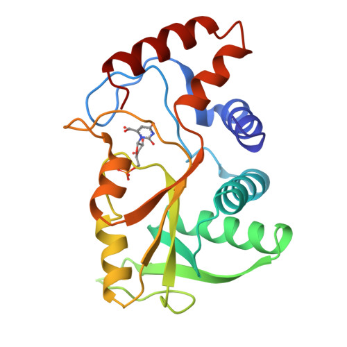
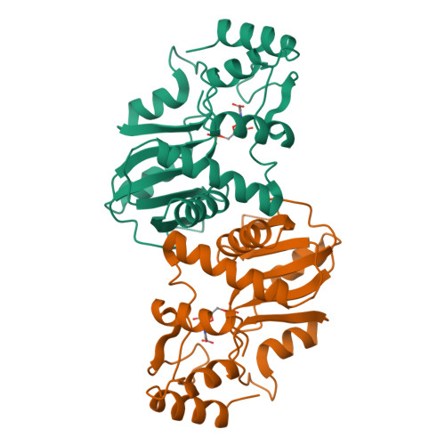
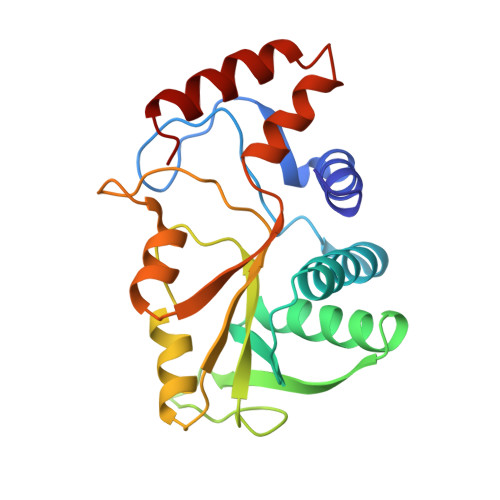
Crystal structure of orotate phosphoribosyltransferase.

Scapin, G.Grubmeyer, C.Sacchettini, J.C.

(1994) Biochemistry 33: 1287-1294

  • DOI: https://doi.org/10.1021/bi00172a001
  • Primary Citation of Related Structures:  
    1STO

  • PubMed Abstract: 

    Phosphoribosyltransferases (PRTases) are enzymes involved in the synthesis of purine, pyrimidine, and pyridine nucleotides. They utilize alpha-D-5-phosphoribosyl-1-pyrophosphate (PRPP) and a nitrogenous base to form a beta-N-riboside monophosphate and pyrophosphate (PPi), and their functional significance in nucleotide homeostasis is evidenced by the devastating effects of inherited diseases associated with the decreased activity and/or stability of these enzymes. The 2.6-A structure of the Salmonella typhimurium orotate phosphoribosyltransferase (OPRTase) complexed with its product orotidine monophosphate (OMP) provides the first detailed image of a member of this group of enzymes. The OPRTase three-dimensional structure was solved using multiple isomorphous replacement methods and reveals two major features: a core five-stranded alpha/beta twisted sheet and an N-terminal region that partially covers the C-terminal portion of the core. PRTases show a very high degree of base specificity. In OPRTase, this is determined by steric constraints and the position of hydrogen bond donors/acceptors of a solvent-inaccessible crevice where the orotate ring of bound OMP resides. Crystalline OPRTase is a dimer, with catalytically important residues from each subunit available to the neighboring subunit, suggesting that oligomerization is necessary for its activity. On the basis of the presence of a common PRPP binding motif among PRTases and the similar chemistry these enzymes perform, we propose that the alpha/beta core found in OPRTase will represent a common feature for PRTases. This generality is demonstrated by construction of a model of the human hypoxanthine-guanine phosphoribosyltransferase (HGPRTase) from secondary structure predictions for HGPRTase and the three-dimensional structure of OPRTase.


  • Organizational Affiliation

    Department of Biochemistry, Albert Einstein College of Medicine, Bronx, New York 10461.


Macromolecules
Find similar proteins by:  (by identity cutoff)  |  3D Structure
Entity ID: 1
MoleculeChains Sequence LengthOrganismDetailsImage
OROTATE PHOSPHORIBOSYLTRANSFERASE213Salmonella enterica subsp. enterica serovar TyphimuriumMutation(s): 0 
EC: 2.4.2.10
UniProt
Find proteins for P08870 (Salmonella typhimurium (strain LT2 / SGSC1412 / ATCC 700720))
Explore P08870 
Go to UniProtKB:  P08870
Entity Groups  
Sequence Clusters30% Identity50% Identity70% Identity90% Identity95% Identity100% Identity
UniProt GroupP08870
Sequence Annotations
Expand
  • Reference Sequence
Small Molecules
Ligands 1 Unique
IDChains Name / Formula / InChI Key2D Diagram3D Interactions
OMP
Query on OMP

Download Ideal Coordinates CCD File 
B [auth A]OROTIDINE-5'-MONOPHOSPHATE
C10 H13 N2 O11 P
KYOBSHFOBAOFBF-XVFCMESISA-N
Experimental Data & Validation

Experimental Data

  • Method: X-RAY DIFFRACTION
  • Resolution: 2.60 Å
  • R-Value Work:  0.176 (Depositor) 
  • R-Value Observed: 0.176 (Depositor) 
Space Group: P 41 21 2
Unit Cell:
Length ( Å )Angle ( ˚ )
a = 47.2α = 90
b = 47.2β = 90
c = 216.68γ = 90
Software Package:
Software NamePurpose
X-PLORmodel building
TNTrefinement
X-PLORrefinement
X-PLORphasing

Structure Validation

View Full Validation Report


Entry History 

Deposition Data

Revision History  (Full details and data files)

  • Version 1.0: 1994-05-31
    Type: Initial release
  • Version 1.1: 2008-03-24
    Changes: Version format compliance
  • Version 1.2: 2011-07-13
    Changes: Derived calculations, Version format compliance
  • Version 1.3: 2019-07-17
    Changes: Data collection, Derived calculations, Other, Refinement description
  • Version 1.4: 2019-08-14
    Changes: Data collection, Refinement description
  • Version 2.0: 2024-09-25
    Changes: Advisory, Atomic model, Data collection, Database references, Derived calculations, Non-polymer description, Structure summary