RCSB PDB - 1QY4: Crystal structure of phosphoglucose isomerase from Pyrococcus furiosus in complex with gluconate 6-phosphate

 1QY4

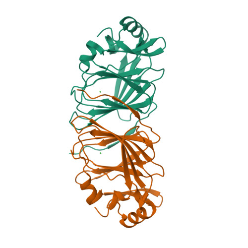
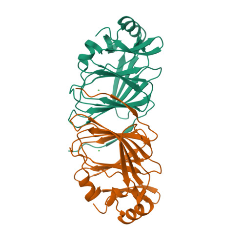
Crystal structure of phosphoglucose isomerase from Pyrococcus furiosus in complex with gluconate 6-phosphate


Experimental Data Snapshot

  • Method: X-RAY DIFFRACTION
  • Resolution: 1.80 Å
  • R-Value Free: 0.266 
  • R-Value Work: 0.218 
  • R-Value Observed: 0.220 

Starting Model: experimental
View more details

wwPDB Validation   3D Report Full Report


Ligand Structure Quality Assessment 

Created with Raphaël 2.3.0Worse 01 BetterLigand structure goodness of fit to experimental dataBest fitted 6PGClick on this verticalbar to view details

This is version 1.3 of the entry. See complete history


Literature

Structural evidence for a hydride transfer mechanism of catalysis in phosphoglucose isomerase from Pyrococcus furiosus

Swan, M.K.Solomons, J.T.G.Beeson, C.C.Hansen, T.Schonheit, P.Davies, C.

(2003) J Biol Chem 278: 47261-47268

  • DOI: https://doi.org/10.1074/jbc.M308603200
  • Primary Citation of Related Structures:  
    1QXJ, 1QXR, 1QY4

  • PubMed Abstract: 

    In the Euryarchaeota species Pyrococcus furiosus and Thermococcus litoralis, phosphoglucose isomerase (PGI) activity is catalyzed by an enzyme unrelated to the well known family of PGI enzymes found in prokaryotes, eukaryotes, and some archaea. We have determined the crystal structure of PGI from Pyrococcus furiosus in native form and in complex with two active site ligands, 5-phosphoarabinonate and gluconate 6-phosphate. In these structures, the metal ion, which in vivo is presumed to be Fe2+, is located in the core of the cupin fold and is immediately adjacent to the C1-C2 region of the ligands, suggesting that Fe2+ is involved in catalysis rather than serving a structural role. The active site contains a glutamate residue that contacts the substrate, but, because it is also coordinated to the metal ion, it is highly unlikely to mediate proton transfer in a cis-enediol mechanism. Consequently, we propose a hydride shift mechanism of catalysis. In this mechanism, Fe2+ is responsible for proton transfer between O1 and O2, and the hydride shift between C1 and C2 is favored by a markedly hydrophobic environment in the active site. The absence of any obvious enzymatic machinery for catalyzing ring opening of the sugar substrates suggests that pyrococcal PGI has a preference for straight chain substrates and that metabolism in extreme thermophiles may use sugars in both ring and straight chain forms.


  • Organizational Affiliation

    Department of Biochemistry and Molecular Biology, Medical University of South Carolina, 173 Ashley Avenue, Charleston, SC 29425, USA.


Macromolecules
Find similar proteins by:  (by identity cutoff)  |  3D Structure
Entity ID: 1
MoleculeChains Sequence LengthOrganismDetailsImage
Glucose-6-phosphate isomerase
A, B
189Pyrococcus furiosusMutation(s): 0 
Gene Names: PGIA OR PF0196
EC: 5.3.1.9
UniProt
Find proteins for P83194 (Pyrococcus furiosus (strain ATCC 43587 / DSM 3638 / JCM 8422 / Vc1))
Explore P83194 
Go to UniProtKB:  P83194
Entity Groups  
Sequence Clusters30% Identity50% Identity70% Identity90% Identity95% Identity100% Identity
UniProt GroupP83194
Sequence Annotations
Expand
  • Reference Sequence
Experimental Data & Validation

Experimental Data

  • Method: X-RAY DIFFRACTION
  • Resolution: 1.80 Å
  • R-Value Free: 0.266 
  • R-Value Work: 0.218 
  • R-Value Observed: 0.220 
  • Space Group: P 1
Unit Cell:
Length ( Å )Angle ( ˚ )
a = 36α = 73.7
b = 39.7β = 78.5
c = 74.7γ = 72.6
Software Package:
Software NamePurpose
REFMACrefinement
CrystalCleardata reduction
CrystalCleardata scaling

Structure Validation

View Full Validation Report


Ligand Structure Quality Assessment 

Created with Raphaël 2.3.0Worse 01 BetterLigand structure goodness of fit to experimental dataBest fitted 6PGClick on this verticalbar to view details

Entry History 

Deposition Data

Revision History  (Full details and data files)

  • Version 1.0: 2003-12-09
    Type: Initial release
  • Version 1.1: 2008-04-29
    Changes: Version format compliance
  • Version 1.2: 2011-07-13
    Changes: Version format compliance
  • Version 1.3: 2023-08-23
    Changes: Advisory, Data collection, Database references, Derived calculations, Refinement description