1NF6

X-ray structure of the Desulfovibrio desulfuricans bacterioferritin: the diiron site in different catalytic states ("cycled" structure: reduced in solution and allowed to reoxidise before crystallisation)


Experimental Data Snapshot

  • Method: X-RAY DIFFRACTION
  • Resolution: 2.35 Å
  • R-Value Free: 0.267 
  • R-Value Work: 0.197 
  • R-Value Observed: 0.197 

Starting Model: experimental
View more details

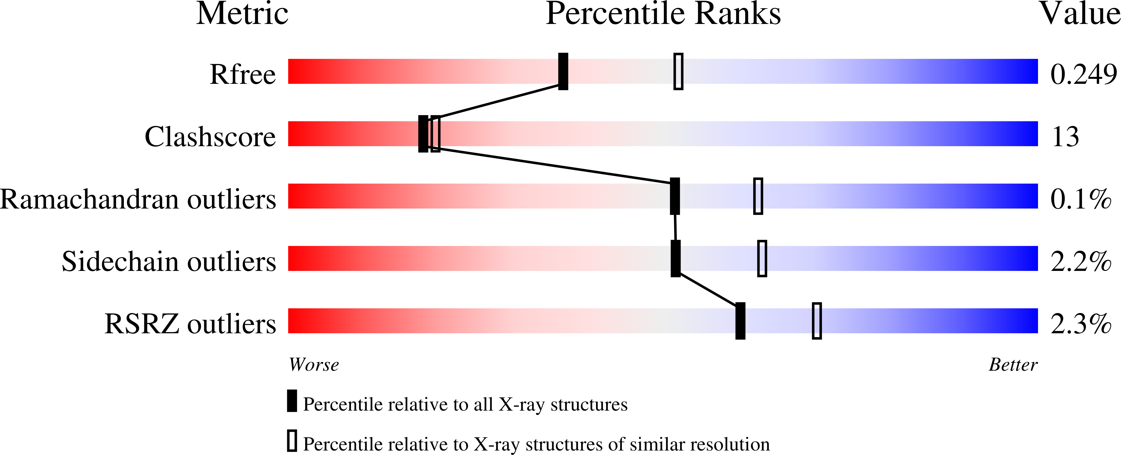
wwPDB Validation   3D Report Full Report


Ligand Structure Quality Assessment 


This is version 1.5 of the entry. See complete history


Literature

The nature of the di-iron site in the bacterioferritin from Desulfovibrio desulfuricans

Macedo, S.Romao, C.V.Mitchell, E.Matias, P.M.Liu, M.Y.Xavier, A.V.LeGall, J.Teixeira, M.Lindley, P.Carrondo, M.A.

(2003) Nat Struct Biol 10: 285-290

  • DOI: https://doi.org/10.1038/nsb909
  • Primary Citation of Related Structures:  
    1NF4, 1NF6, 1NFV

  • PubMed Abstract: 

    The first crystal structure of a native di-iron center in an iron-storage protein (bacterio)ferritin is reported. The protein, isolated from the anaerobic bacterium Desulfovibrio desulfuricans, has the unique property of having Fe-coproporphyrin III as its heme cofactor. The three-dimensional structure of this bacterioferritin was determined in three distinct catalytic/redox states by X-ray crystallography (at 1.95, 2.05 and 2.35 A resolution), corresponding to different intermediates of the di-iron ferroxidase site. Conformational changes associated with these intermediates support the idea of a route for iron entry into the protein shell through a pore that passes through the di-iron center. Molecular surface and electrostatic potential calculations also suggest the presence of another ion channel, distant from the channels at the three- and four-fold axes proposed as points of entry for the iron atoms.


  • Organizational Affiliation

    Instituto de Tecnologia Química e Biológica, Universidade Nova de Lisboa, Av. República, EAN, Apartado 127, 2781-901 Oeiras, Portugal.


Macromolecules
Find similar proteins by:  (by identity cutoff)  |  3D Structure
Entity ID: 1
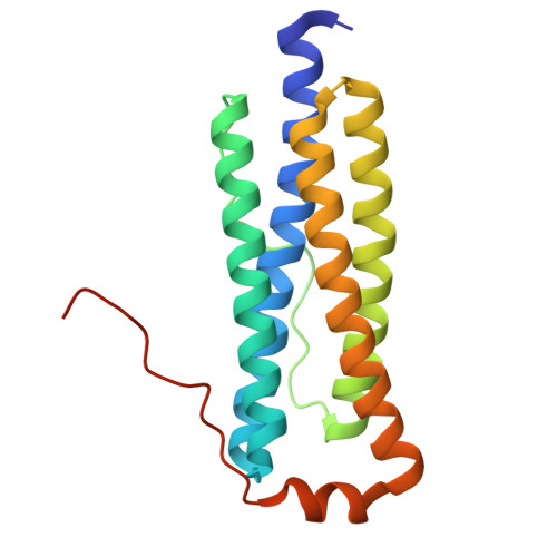
MoleculeChains Sequence LengthOrganismDetailsImage
bacterioferritin
A, B, C, D, E
A, B, C, D, E, F, G, H, I, J, K, L, M, N, O, P
179Desulfovibrio desulfuricansMutation(s): 0 
EC: 1.16.3.1
UniProt
Find proteins for Q93PP9 (Desulfovibrio desulfuricans (strain ATCC 27774 / DSM 6949 / MB))
Explore Q93PP9 
Go to UniProtKB:  Q93PP9
Entity Groups  
Sequence Clusters30% Identity50% Identity70% Identity90% Identity95% Identity100% Identity
UniProt GroupQ93PP9
Sequence Annotations
Expand
  • Reference Sequence
Small Molecules
Ligands 4 Unique
IDChains Name / Formula / InChI Key2D Diagram3D Interactions
FEC
Query on FEC

Download Ideal Coordinates CCD File 
AA [auth C]
EB [auth K]
JA [auth F]
NA [auth G]
QB [auth N]
AA [auth C],
EB [auth K],
JA [auth F],
NA [auth G],
QB [auth N],
TB [auth O],
U [auth A],
VA [auth I]
1,3,5,8-TETRAMETHYL-PORPHINE-2,4,6,7-TETRAPROPIONIC ACID FERROUS COMPLEX
C36 H36 Fe N4 O8
FEDZMOFKVKOYTI-RGGAHWMASA-L
SO4
Query on SO4

Download Ideal Coordinates CCD File 
BB [auth K]
CA [auth D]
CB [auth K]
DA [auth E]
DB [auth K]
BB [auth K],
CA [auth D],
CB [auth K],
DA [auth E],
DB [auth K],
FA [auth E],
GB [auth L],
HA [auth F],
HB [auth L],
IA [auth F],
JB [auth M],
KB [auth M],
LA [auth G],
LB [auth M],
MA [auth G],
MB [auth N],
OB [auth N],
PA [auth H],
PB [auth N],
QA [auth H],
R [auth A],
RA [auth I],
S [auth A],
SB [auth O],
T [auth A],
TA [auth I],
UA [auth I],
VB [auth P],
W [auth B],
XA [auth J],
Y [auth C],
YA [auth J],
Z [auth C],
ZA [auth K]
SULFATE ION
O4 S
QAOWNCQODCNURD-UHFFFAOYSA-L
GOL
Query on GOL

Download Ideal Coordinates CCD File 
WB [auth P]GLYCEROL
C3 H8 O3
PEDCQBHIVMGVHV-UHFFFAOYSA-N
FE
Query on FE

Download Ideal Coordinates CCD File 
AB [auth K]
BA [auth D]
EA [auth E]
FB [auth L]
GA [auth F]
AB [auth K],
BA [auth D],
EA [auth E],
FB [auth L],
GA [auth F],
IB [auth M],
KA [auth G],
NB [auth N],
OA [auth H],
Q [auth A],
RB [auth O],
SA [auth I],
UB [auth P],
V [auth B],
WA [auth J],
X [auth C]
FE (III) ION
Fe
VTLYFUHAOXGGBS-UHFFFAOYSA-N
Experimental Data & Validation

Experimental Data

  • Method: X-RAY DIFFRACTION
  • Resolution: 2.35 Å
  • R-Value Free: 0.267 
  • R-Value Work: 0.197 
  • R-Value Observed: 0.197 
  • Space Group: P 21 3
Unit Cell:
Length ( Å )Angle ( ˚ )
a = 225.79α = 90
b = 225.79β = 90
c = 225.79γ = 90
Software Package:
Software NamePurpose
SHELXL-97refinement
SCALAdata scaling
CNSrefinement
DENZOdata reduction
CCP4data scaling
CNSphasing

Structure Validation

View Full Validation Report


Ligand Structure Quality Assessment 


Entry History 

Deposition Data

Revision History  (Full details and data files)

  • Version 1.0: 2003-04-01
    Type: Initial release
  • Version 1.1: 2008-04-29
    Changes: Version format compliance
  • Version 1.2: 2011-07-13
    Changes: Derived calculations, Version format compliance
  • Version 1.3: 2019-07-24
    Changes: Advisory, Data collection, Derived calculations, Refinement description
  • Version 1.4: 2024-04-03
    Changes: Advisory, Data collection, Database references, Derived calculations, Refinement description
  • Version 1.5: 2024-12-25
    Changes: Advisory, Derived calculations, Structure summary