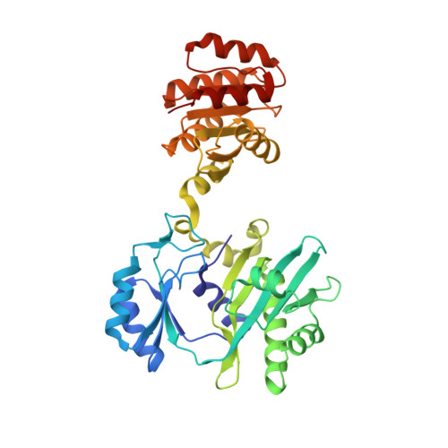
1JLL

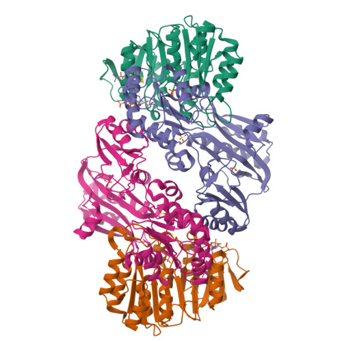
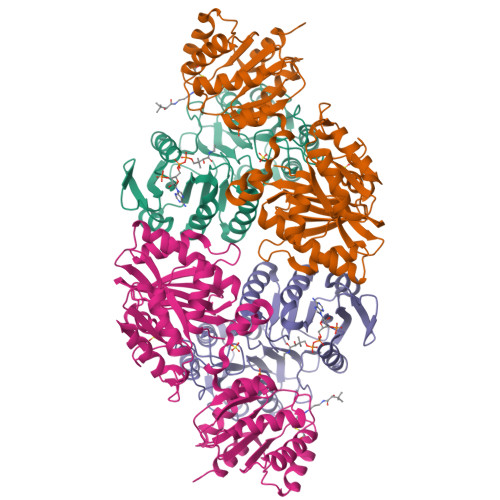
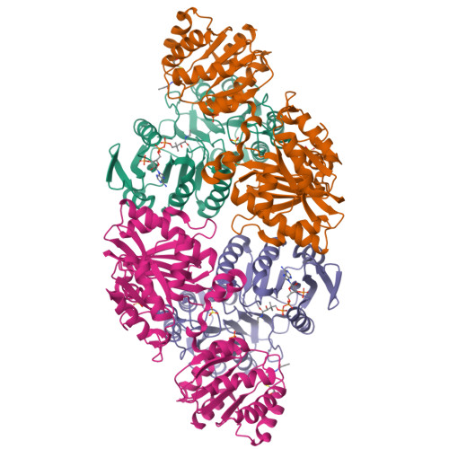
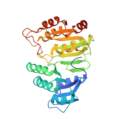
Crystal Structure Analysis of the E197betaA Mutant of E. coli SCS


Experimental Data Snapshot

  • Method: X-RAY DIFFRACTION
  • Resolution: 2.69 Å
  • R-Value Free: 0.259 
  • R-Value Work: 0.222 
  • R-Value Observed: 0.223 

Starting Model: experimental
View more details

wwPDB Validation   3D Report Full Report


Ligand Structure Quality Assessment 


This is version 1.5 of the entry. See complete history


Literature

Two glutamate residues, Glu 208 alpha and Glu 197 beta, are crucial for phosphorylation and dephosphorylation of the active-site histidine residue in succinyl-CoA synthetase.

Fraser, M.E.Joyce, M.A.Ryan, D.G.Wolodko, W.T.

(2002) Biochemistry 41: 537-546

  • DOI: https://doi.org/10.1021/bi011518y
  • Primary Citation of Related Structures:  
    1JKJ, 1JLL

  • PubMed Abstract: 

    Succinyl-CoA synthetase catalyzes the reversible reaction succinyl-CoA + NDP + P(i) <--> succinate + CoA + NTP (N denoting adenosine or guanosine). The enzyme consists of two different subunits, designated alpha and beta. During the reaction, a histidine residue of the alpha-subunit is transiently phosphorylated. This histidine residue interacts with Glu 208 alpha at site I in the structures of phosphorylated and dephosphorylated Escherichia coli SCS. We postulated that Glu 197 beta, a residue in the nucleotide-binding domain, would provide similar stabilization of the histidine residue during the actual phosphorylation/dephosphorylation by nucleotide at site II. In this work, these two glutamate residues have been mutated individually to aspartate or glutamine. Glu 197 beta has been additionally mutated to alanine. The mutant proteins were tested for their ability to be phosphorylated in the forward or reverse direction. The aspartate mutant proteins can be phosphorylated in either direction, while the E208 alpha Q mutant protein can only be phosphorylated by NTP, and the E197 beta Q mutant protein can only be phosphorylated by succinyl-CoA and P(i). These results demonstrate that the length of the side chain at these positions is not critical, but that the charge is. Most significantly, the E197 beta A mutant protein could not be phosphorylated in either direction. Its crystal structure shows large differences from the wild-type enzyme in the conformation of two residues of the alpha-subunit, Cys 123 alpha-Pro 124 alpha. We postulate that in this conformation, the protein cannot productively bind succinyl-CoA for phosphorylation via succinyl-CoA and P(i).


  • Organizational Affiliation

    Department of Biochemistry, University of Western Ontario, London, Ontario, Canada N6A 5C1. [email protected]


Macromolecules
Find similar proteins by:  (by identity cutoff)  |  3D Structure
Entity ID: 1
MoleculeChains Sequence LengthOrganismDetailsImage
succinyl-CoA synthetase alpha subunitA,
C [auth D]
288Escherichia coliMutation(s): 0 
EC: 6.2.1.5
UniProt
Find proteins for P0AGE9 (Escherichia coli (strain K12))
Explore P0AGE9 
Go to UniProtKB:  P0AGE9
Entity Groups  
Sequence Clusters30% Identity50% Identity70% Identity90% Identity95% Identity100% Identity
UniProt GroupP0AGE9
Sequence Annotations
Expand
  • Reference Sequence
Find similar proteins by:  (by identity cutoff)  |  3D Structure
Entity ID: 2
MoleculeChains Sequence LengthOrganismDetailsImage
succinyl-CoA synthetase beta subunitB,
D [auth E]
388Escherichia coliMutation(s): 1 
EC: 6.2.1.5
UniProt
Find proteins for P0A836 (Escherichia coli (strain K12))
Explore P0A836 
Go to UniProtKB:  P0A836
Entity Groups  
Sequence Clusters30% Identity50% Identity70% Identity90% Identity95% Identity100% Identity
UniProt GroupP0A836
Sequence Annotations
Expand
  • Reference Sequence
Small Molecules
Ligands 3 Unique
IDChains Name / Formula / InChI Key2D Diagram3D Interactions
COA
Query on COA

Download Ideal Coordinates CCD File 
F [auth A],
I [auth B],
K [auth D],
N [auth E]
COENZYME A
C21 H36 N7 O16 P3 S
RGJOEKWQDUBAIZ-IBOSZNHHSA-N
SO4
Query on SO4

Download Ideal Coordinates CCD File 
G [auth B],
H [auth B],
L [auth E],
M [auth E]
SULFATE ION
O4 S
QAOWNCQODCNURD-UHFFFAOYSA-L
PO4
Query on PO4

Download Ideal Coordinates CCD File 
E [auth A],
J [auth D]
PHOSPHATE ION
O4 P
NBIIXXVUZAFLBC-UHFFFAOYSA-K
Experimental Data & Validation

Experimental Data

  • Method: X-RAY DIFFRACTION
  • Resolution: 2.69 Å
  • R-Value Free: 0.259 
  • R-Value Work: 0.222 
  • R-Value Observed: 0.223 
  • Space Group: P 43 2 2
Unit Cell:
Length ( Å )Angle ( ˚ )
a = 97.06α = 90
b = 97.06β = 90
c = 389.6γ = 90
Software Package:
Software NamePurpose
CNSrefinement
DENZOdata reduction
SCALEPACKdata scaling
CNSphasing

Structure Validation

View Full Validation Report


Ligand Structure Quality Assessment 


Entry History 

Deposition Data

  • Released Date: 2002-01-30 
  • Deposition Author(s): Fraser, M.E.

Revision History  (Full details and data files)

  • Version 1.0: 2002-01-30
    Type: Initial release
  • Version 1.1: 2008-04-27
    Changes: Version format compliance
  • Version 1.2: 2011-07-13
    Changes: Version format compliance
  • Version 1.3: 2021-10-27
    Changes: Database references, Derived calculations
  • Version 1.4: 2023-08-16
    Changes: Data collection, Refinement description
  • Version 1.5: 2024-11-06
    Changes: Structure summary