1FOA | pdb_00001foa

CRYSTAL STRUCTURE OF N-ACETYLGLUCOSAMINYLTRANSFERASE I


Experimental Data Snapshot

  • Method: X-RAY DIFFRACTION
  • Resolution: 1.80 Å
  • R-Value Free: 
    0.229 (Depositor), 0.226 (DCC) 
  • R-Value Work: 
    0.185 (Depositor), 0.179 (DCC) 
  • R-Value Observed: 
    0.185 (Depositor) 

wwPDB Validation   3D Report Full Report


Ligand Structure Quality Assessment 

Created with Raphaël 2.3.0Worse 01 BetterLigand structure goodness of fit to experimental dataBest fitted UD1Click on this verticalbar to view details

This is version 1.3 of the entry. See complete history


Literature

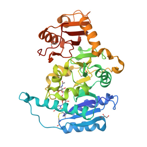
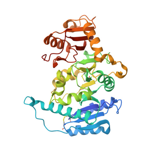
X-ray crystal structure of rabbit N-acetylglucosaminyltransferase I: catalytic mechanism and a new protein superfamily.

Unligil, U.M.Zhou, S.Yuwaraj, S.Sarkar, M.Schachter, H.Rini, J.M.

(2000) EMBO J 19: 5269-5280

  • DOI: https://doi.org/10.1093/emboj/19.20.5269
  • Primary Citation of Related Structures:  
    1FO8, 1FO9, 1FOA

  • PubMed Abstract: 

    N:-acetylglucosaminyltransferase I (GnT I) serves as the gateway from oligomannose to hybrid and complex N:-glycans and plays a critical role in mammalian development and possibly all metazoans. We have determined the X-ray crystal structure of the catalytic fragment of GnT I in the absence and presence of bound UDP-GlcNAc/Mn(2+) at 1.5 and 1.8 A resolution, respectively. The structures identify residues critical for substrate binding and catalysis and provide evidence for similarity, at the mechanistic level, to the deglycosylation step of retaining beta-glycosidases. The structuring of a 13 residue loop, resulting from UDP-GlcNAc/Mn(2+) binding, provides an explanation for the ordered sequential 'Bi Bi' kinetics shown by GnT I. Analysis reveals a domain shared with Bacillus subtilis glycosyltransferase SpsA, bovine beta-1,4-galactosyl transferase 1 and Escherichia coli N:-acetylglucosamine-1-phosphate uridyltransferase. The low sequence identity, conserved fold and related functional features shown by this domain define a superfamily whose members probably share a common ancestor. Sequence analysis and protein threading show that the domain is represented in proteins from several glycosyltransferase families.


  • Organizational Affiliation

    Department of Medical Genetics and Microbiology, University of Toronto, Toronto, Ontario M5S 1A8, Canada.


Macromolecules
Find similar proteins by:  (by identity cutoff)  |  3D Structure
Entity ID: 1
MoleculeChains Sequence LengthOrganismDetailsImage
ALPHA-1,3-MANNOSYL-GLYCOPROTEIN BETA-1,2-N-ACETYLGLUCOSAMINYLTRANSFERASE348Oryctolagus cuniculusMutation(s): 0 
EC: 2.4.1.101
UniProt
Find proteins for P27115 (Oryctolagus cuniculus)
Explore P27115 
Go to UniProtKB:  P27115
Entity Groups  
Sequence Clusters30% Identity50% Identity70% Identity90% Identity95% Identity100% Identity
UniProt GroupP27115
Sequence Annotations
Expand
  • Reference Sequence
Experimental Data & Validation

Experimental Data

  • Method: X-RAY DIFFRACTION
  • Resolution: 1.80 Å
  • R-Value Free:  0.229 (Depositor), 0.226 (DCC) 
  • R-Value Work:  0.185 (Depositor), 0.179 (DCC) 
  • R-Value Observed: 0.185 (Depositor) 
Space Group: P 21 21 21
Unit Cell:
Length ( Å )Angle ( ˚ )
a = 40.541α = 90
b = 82.19β = 90
c = 101.956γ = 90
Software Package:
Software NamePurpose
DENZOdata reduction
SCALEPACKdata scaling
SOLVEphasing
CNSrefinement

Structure Validation

View Full Validation Report


Ligand Structure Quality Assessment 

Created with Raphaël 2.3.0Worse 01 BetterLigand structure goodness of fit to experimental dataBest fitted UD1Click on this verticalbar to view details

Entry History 

Deposition Data

Revision History  (Full details and data files)

  • Version 1.0: 2001-05-16
    Type: Initial release
  • Version 1.1: 2008-04-27
    Changes: Version format compliance
  • Version 1.2: 2011-07-13
    Changes: Non-polymer description, Version format compliance
  • Version 1.3: 2024-11-20
    Changes: Data collection, Database references, Derived calculations, Structure summary