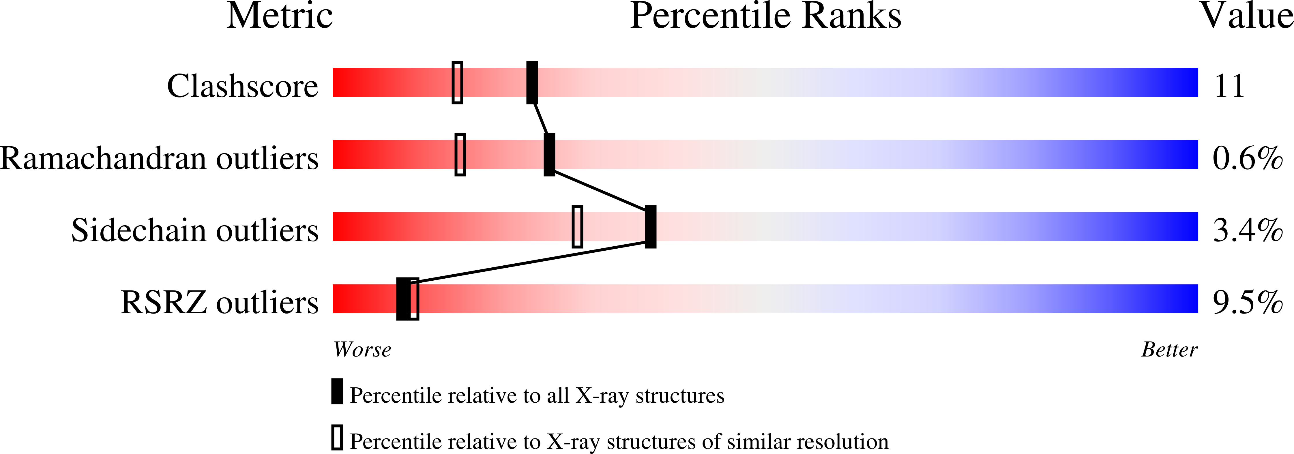
1DMK

BOVINE ENDOTHELIAL NITRIC OXIDE SYNTHASE HEME DOMAIN COMPLEXED WITH 4-AMINO-6-PHENYL-TETRAHYDROPTERIDINE


Experimental Data Snapshot

  • Method: X-RAY DIFFRACTION
  • Resolution: 1.90 Å
  • R-Value Free: 0.264 
  • R-Value Work: 0.225 
  • R-Value Observed: 0.225 

wwPDB Validation   3D Report Full Report


Ligand Structure Quality Assessment 


This is version 1.3 of the entry. See complete history


Literature

Structural basis for pterin antagonism in nitric-oxide synthase. Development of novel 4-oxo-pteridine antagonists of (6R)-5,6,7,8-tetrahydrobiopterin

Kotsonis, P.Frohlich, L.G.Raman, C.S.Li, H.Berg, M.Gerwig, R.Groehn, V.Kang, Y.Al-Masoudi, N.Taghavi-Moghadam, S.Mohr, D.Munch, U.Schnabel, J.Martasek, P.Masters, B.S.Strobel, H.Poulos, T.Matter, H.Pfleiderer, W.Schmidt, H.H.

(2001) J Biol Chem 276: 49133-49141

  • DOI: https://doi.org/10.1074/jbc.M011469200
  • Primary Citation of Related Structures:  
    1DMJ, 1DMK

  • PubMed Abstract: 

    Pathological nitric oxide (NO) generation in sepsis, inflammation, and stroke may be therapeutically controlled by inhibiting NO synthases (NOS). Here we targeted the (6R)-5,6,7,8-tetrahydro-l-biopterin (H(4)Bip)-binding site of NOS, which, upon cofactor binding, maximally increases enzyme activity and NO production from substrate l-arginine. The first generation of H(4)Bip-based NOS inhibitors employed a 4-amino pharmacophore of H(4)Bip analogous to antifolates such as methotrexate. We developed a novel series of 4-oxo-pteridine derivatives that were screened for inhibition against neuronal NOS (NOS-I) and a structure-activity relationship was determined. To understand the structural basis for pterin antagonism, selected derivatives were docked into the NOS pterin binding cavity. Using a reduced 4-oxo-pteridine scaffold, derivatives with certain modifications such as electron-rich aromatic phenyl or benzoyl groups at the 5- and 6-positions, were discovered to markedly inhibit NOS-I, possibly due to hydrophobic and electrostatic interactions with Phe(462) and Ser(104), respectively, within the pterin binding pocket. One of the most effective 4-oxo compounds and, for comparisons an active 4-amino derivative, were then co-crystallized with the endothelial NOS (NOS-III) oxygenase domain and this structure solved to confirm the hypothetical binding modes. Collectively, these findings suggest (i) that, unlike the antifolate principle, the 4-amino substituent is not essential for developing pterin-based NOS inhibitors and (ii), provide a steric and electrostatic basis for their rational design.


  • Organizational Affiliation

    Department of Pharmacology and Toxicology, Julius-Maximilians University, Versbacher Strasse 9, Würzburg 97078, Germany. [email protected]


Macromolecules
Find similar proteins by:  (by identity cutoff)  |  3D Structure
Entity ID: 1
MoleculeChains Sequence LengthOrganismDetailsImage
NITRIC OXIDE SYNTHASE
A, B
444Bos taurusMutation(s): 0 
EC: 1.14.13.39
UniProt
Find proteins for P29473 (Bos taurus)
Explore P29473 
Go to UniProtKB:  P29473
Entity Groups  
Sequence Clusters30% Identity50% Identity70% Identity90% Identity95% Identity100% Identity
UniProt GroupP29473
Sequence Annotations
Expand
  • Reference Sequence
Small Molecules
Ligands 6 Unique
IDChains Name / Formula / InChI Key2D Diagram3D Interactions
HEM
Query on HEM

Download Ideal Coordinates CCD File 
H [auth A],
P [auth B]
PROTOPORPHYRIN IX CONTAINING FE
C34 H32 Fe N4 O4
KABFMIBPWCXCRK-RGGAHWMASA-L
AP6
Query on AP6

Download Ideal Coordinates CCD File 
I [auth A],
Q [auth B]
2,4-DIAMINO-6-PHENYL-5,6,7,8,-TETRAHYDROPTERIDINE
C12 H14 N6
VEKRIXRQADJFAG-QMMMGPOBSA-N
CAC
Query on CAC

Download Ideal Coordinates CCD File 
F [auth A],
O [auth B]
CACODYLATE ION
C2 H6 As O2
OGGXGZAMXPVRFZ-UHFFFAOYSA-M
ITU
Query on ITU

Download Ideal Coordinates CCD File 
J [auth A],
R [auth B]
ETHYLISOTHIOUREA
C3 H8 N2 S
VFIZBHJTOHUOEK-UHFFFAOYSA-N
ZN
Query on ZN

Download Ideal Coordinates CCD File 
G [auth A]ZINC ION
Zn
PTFCDOFLOPIGGS-UHFFFAOYSA-N
ACT
Query on ACT

Download Ideal Coordinates CCD File 
C [auth A]
D [auth A]
E [auth A]
K [auth B]
L [auth B]
C [auth A],
D [auth A],
E [auth A],
K [auth B],
L [auth B],
M [auth B],
N [auth B]
ACETATE ION
C2 H3 O2
QTBSBXVTEAMEQO-UHFFFAOYSA-M
Binding Affinity Annotations 
IDSourceBinding Affinity
ITU BindingDB:  1DMK Ki: 39 (nM) from 1 assay(s)
IC50: 1300 (nM) from 1 assay(s)
AP6 BindingDB:  1DMK IC50: 6000 (nM) from 1 assay(s)
Experimental Data & Validation

Experimental Data

  • Method: X-RAY DIFFRACTION
  • Resolution: 1.90 Å
  • R-Value Free: 0.264 
  • R-Value Work: 0.225 
  • R-Value Observed: 0.225 
  • Space Group: P 21 21 21
Unit Cell:
Length ( Å )Angle ( ˚ )
a = 58.07α = 90
b = 106.11β = 90
c = 156.21γ = 90
Software Package:
Software NamePurpose
X-PLORmodel building
CNSrefinement
DENZOdata reduction
SCALEPACKdata scaling
X-PLORphasing

Structure Validation

View Full Validation Report


Ligand Structure Quality Assessment 


Entry History 

Revision History  (Full details and data files)

  • Version 1.0: 2000-12-20
    Type: Initial release
  • Version 1.1: 2008-04-27
    Changes: Version format compliance
  • Version 1.2: 2011-07-13
    Changes: Non-polymer description, Version format compliance
  • Version 1.3: 2024-02-07
    Changes: Data collection, Database references, Derived calculations