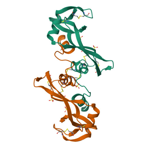
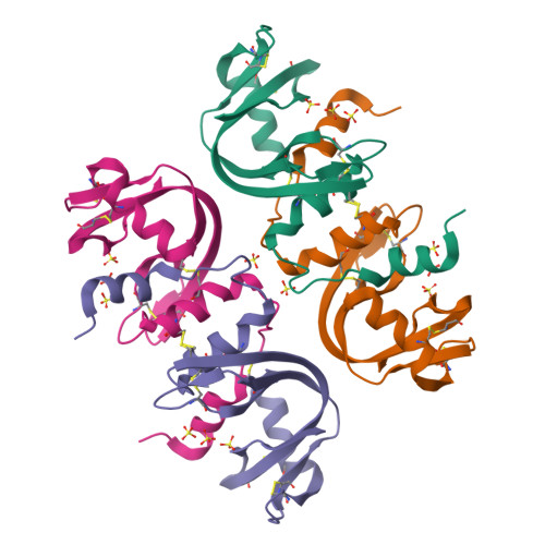
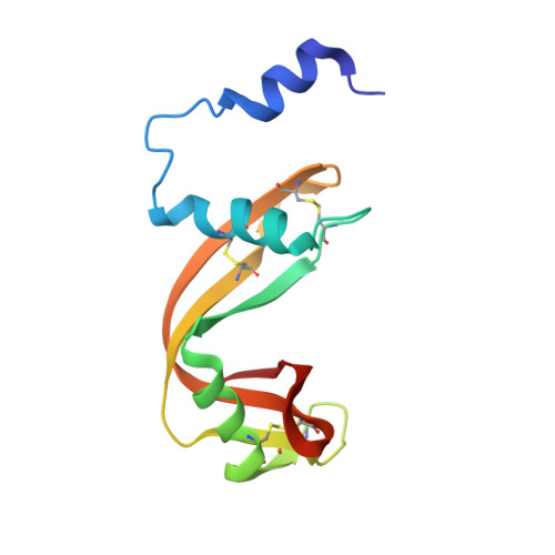
Bovine seminal ribonuclease: structure at 1.9 A resolution.
Mazzarella, L., Capasso, S., Demasi, D., Di Lorenzo, G., Mattia, C.A., Zagari, A.(1993) Acta Crystallogr D Biol Crystallogr 49: 389-402
- PubMed: 15299514
- DOI: https://doi.org/10.1107/S0907444993003403
- Primary Citation of Related Structures:
1BSR - PubMed Abstract:
The crystal structure of bovine seminal ribonuclease, a homodimeric enzyme closely related to pancreatic ribonuclease, has been refined at a nominal resolution of 1.9 A employing data collected on an electronic area detector. The final model consists of two chains containing 1990 non-H atoms, seven sulfate anions and 113 water molecules per asymmetric unit. The unit-cell parameters are a = 36.5 (1), b = 66.7 (1) and c = 107.5 (2) A, space group P22(1)2(1). The R factor is 0.177 for 16 492 reflections in the resolution range 6.0-1.9 A and the deviations from ideal values of bond lengths and bond angles are 0.020 A and 3.7 degrees, respectively. The molecule is formed by two pancreatic like chains, which have their N-terminal segments interchanged so that each active site is formed by residues from both subunits. The two chains are related by a non-crystallographic twofold symmetry and are covalently linked by two consecutive disulfide bridges, which form an unusual sixteen-membered ring across the dimer interface. The deviations from the molecular symmetry, the hydration shell and the sulfate-binding sites are also discussed in relation to the known structure of the pancreatic enzyme.
Organizational Affiliation:
Dipartimento di Chimica, Università Federico II, Napoli, Italy.


