9F5H | pdb_00009f5h

Crystal structure of MGAT5 bump-and-hole mutant in complex with UDP and M592


Experimental Data Snapshot

  • Method: X-RAY DIFFRACTION
  • Resolution: 1.97 Å
  • R-Value Free: 
    0.242 (Depositor), 0.242 (DCC) 
  • R-Value Work: 
    0.200 (Depositor), 0.206 (DCC) 

Starting Model: experimental
View more details

wwPDB Validation   3D Report Full Report


Ligand Structure Quality Assessment 

Created with Raphaël 2.3.0Worse 01 BetterLigand structure goodness of fit to experimental dataBest fitted UDPClick on this verticalbar to view details

This is version 1.1 of the entry. See complete history


Literature

A Bioorthogonal Precision Tool for Human N -Acetylglucosaminyltransferase V.

Liu, Y.Bineva-Todd, G.Meek, R.W.Mazo, L.Piniello, B.Moroz, O.Burnap, S.A.Begum, N.Ohara, A.Roustan, C.Tomita, S.Kjaer, S.Polizzi, K.Struwe, W.B.Rovira, C.Davies, G.J.Schumann, B.

(2024) J Am Chem Soc 146: 26707-26718

  • DOI: https://doi.org/10.1021/jacs.4c05955
  • Primary Citation of Related Structures:  
    9F5H

  • PubMed Abstract: 

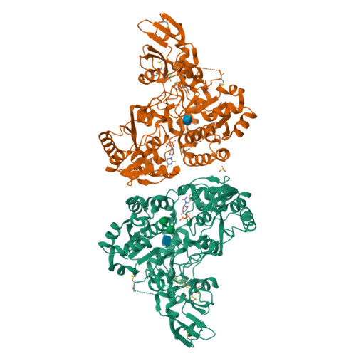
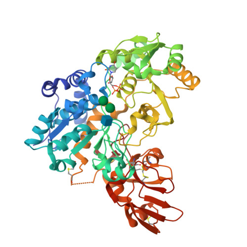
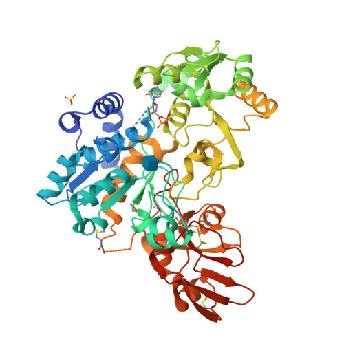
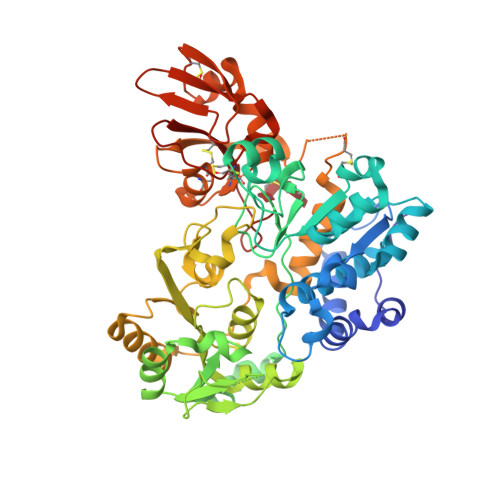
    Correct elaboration of N-linked glycans in the secretory pathway of human cells is essential in physiology. Early N-glycan biosynthesis follows an assembly line principle before undergoing crucial elaboration points that feature the sequential incorporation of the sugar N -acetylglucosamine (GlcNAc). The activity of GlcNAc transferase V (MGAT5) primes the biosynthesis of an N-glycan antenna that is heavily upregulated in cancer. Still, the functional relevance and substrate choice of MGAT5 are ill-defined. Here, we employ protein engineering to develop a bioorthogonal substrate analog for the activity of MGAT5. Chemoenzymatic synthesis is used to produce a collection of nucleotide-sugar analogs with bulky, bioorthogonal acylamide side chains. We find that WT-MGAT5 displays considerable activity toward such substrate analogues. Protein engineering yields an MGAT5 variant that loses activity against the native nucleotide sugar and increases activity toward a 4-azidobutyramide-containing substrate analogue. By such restriction of substrate specificity, we show that the orthogonal enzyme-substrate pair is suitable to bioorthogonally tag glycoproteins. Through X-ray crystallography and molecular dynamics simulations, we establish the structural basis of MGAT5 engineering, informing the design rules for bioorthogonal precision chemical tools.


  • Organizational Affiliation

    Department of Chemistry, Imperial College London, London W12 0BZ, U.K.


Macromolecules
Find similar proteins by:  (by identity cutoff)  |  3D Structure
Entity ID: 1
MoleculeChains Sequence LengthOrganismDetailsImage
Secreted alpha-1,6-mannosylglycoprotein 6-beta-N-acetylglucosaminyltransferase A
A, B
515Homo sapiensMutation(s): 3 
Gene Names: MGAT5GGNT5
EC: 2.4.1.155
UniProt & NIH Common Fund Data Resources
Find proteins for Q09328 (Homo sapiens)
Explore Q09328 
Go to UniProtKB:  Q09328
PHAROS:  Q09328
GTEx:  ENSG00000152127 
Entity Groups  
Sequence Clusters30% Identity50% Identity70% Identity90% Identity95% Identity100% Identity
UniProt GroupQ09328
Sequence Annotations
Expand
  • Reference Sequence
Small Molecules
Ligands 4 Unique
IDChains Name / Formula / InChI Key2D Diagram3D Interactions
UDP (Subject of Investigation/LOI)
Query on UDP

Download Ideal Coordinates CCD File 
C [auth A],
G [auth B]
URIDINE-5'-DIPHOSPHATE
C9 H14 N2 O12 P2
XCCTYIAWTASOJW-XVFCMESISA-N
NAG
Query on NAG

Download Ideal Coordinates CCD File 
D [auth A],
H [auth B]
2-acetamido-2-deoxy-beta-D-glucopyranose
C8 H15 N O6
OVRNDRQMDRJTHS-FMDGEEDCSA-N
MAN
Query on MAN

Download Ideal Coordinates CCD File 
E [auth A],
F [auth A]
alpha-D-mannopyranose
C6 H12 O6
WQZGKKKJIJFFOK-PQMKYFCFSA-N
SO4
Query on SO4

Download Ideal Coordinates CCD File 
I [auth B]SULFATE ION
O4 S
QAOWNCQODCNURD-UHFFFAOYSA-L
Experimental Data & Validation

Experimental Data

  • Method: X-RAY DIFFRACTION
  • Resolution: 1.97 Å
  • R-Value Free:  0.242 (Depositor), 0.242 (DCC) 
  • R-Value Work:  0.200 (Depositor), 0.206 (DCC) 
Space Group: P 1
Unit Cell:
Length ( Å )Angle ( ˚ )
a = 46.507α = 108.208
b = 69.069β = 92.088
c = 90.993γ = 106.747
Software Package:
Software NamePurpose
REFMACrefinement
REFMACrefinement
xia2data reduction
Aimlessdata scaling
MOLREPphasing

Structure Validation

View Full Validation Report


Ligand Structure Quality Assessment 

Created with Raphaël 2.3.0Worse 01 BetterLigand structure goodness of fit to experimental dataBest fitted UDPClick on this verticalbar to view details

Entry History & Funding Information

Deposition Data


Funding OrganizationLocationGrant Number
European Research Council (ERC)European Union951231

Revision History  (Full details and data files)

  • Version 1.0: 2024-10-02
    Type: Initial release
  • Version 1.1: 2024-10-16
    Changes: Database references, Structure summary