1UUM | pdb_00001uum

Rat dihydroorotate dehydrogenase (DHOD)in complex with atovaquone


Experimental Data Snapshot

  • Method: X-RAY DIFFRACTION
  • Resolution: 2.30 Å
  • R-Value Free: 
    0.242 (Depositor), 0.210 (DCC) 
  • R-Value Work: 
    0.216 (Depositor), 0.210 (DCC) 
  • R-Value Observed: 
    0.216 (Depositor) 

wwPDB Validation   3D Report Full Report


Ligand Structure Quality Assessment 

Created with Raphaël 2.3.0Worse 01 BetterLigand structure goodness of fit to experimental dataBest fitted FMNClick on this verticalbar to view detailsBest fitted AFIClick on this verticalbar to view detailsBest fitted BOGClick on this verticalbar to view detailsBest fitted OROClick on this verticalbar to view details

This is version 1.7 of the entry. See complete history


Literature

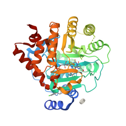
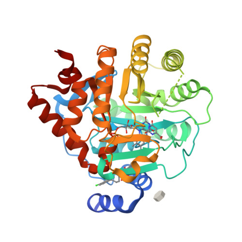
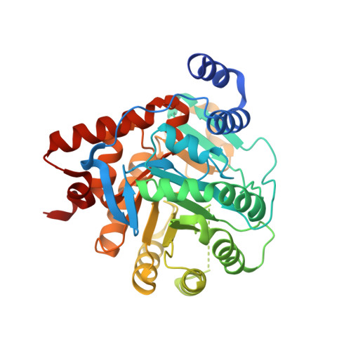
Inhibitor Binding in a Class 2 Dihydroorotate Dehydrogenase Causes Variations in the Membrane-Associated N-Terminal Domain

Hansen, M.Le Nours, J.Johansson, E.Antal, T.Ullrich, A.Loffler, M.Larsen, S.

(2004) Protein Sci 13: 1031

  • DOI: https://doi.org/10.1110/ps.03533004
  • Primary Citation of Related Structures:  
    1UUM, 1UUO

  • PubMed Abstract: 

    The flavin enzyme dihydroorotate dehydrogenase (DHOD; EC 1.3.99.11) catalyzes the oxidation of dihydroorotate to orotate, the fourth step in the de novo pyrimidine biosynthesis of UMP. The enzyme is a promising target for drug design in different biological and clinical applications for cancer and arthritis. The first crystal structure of the class 2 dihydroorotate dehydrogenase from rat has been determined in complex with its two inhibitors brequinar and atovaquone. These inhibitors have shown promising results as anti-proliferative, immunosuppressive, and antiparasitic agents. A unique feature of the class 2 DHODs is their N-terminal extension, which folds into a separate domain comprising two alpha-helices. This domain serves as the binding site for the two inhibitors and the respiratory quinones acting as the second substrate for the class 2 DHODs. The orientation of the first N-terminal helix is very different in the two complexes of rat DHOD (DHODR). Binding of atovaquone causes a 12 A movement of the first residue in the first alpha-helix. Based on the information from the two structures of DHODR, a model for binding of the quinone and the residues important for the interactions could be defined. His 56 and Arg 136, which are fully conserved in all class 2 DHODs, seem to play a key role in the interaction with the electron acceptor. The differences between the membrane-bound rat DHOD and membrane-associated class 2 DHODs exemplified by the Escherichia coli DHOD has been investigated by GRID computations of the hydrophobic probes predicted to interact with the membrane.


  • Organizational Affiliation

    Centre for Crystallographic Studies, Department of Chemistry, University of Copenhagen, Universitetsparken 5, DK-2100 Copenhagen, Denmark.


Macromolecules
Find similar proteins by:  (by identity cutoff)  |  3D Structure
Entity ID: 1
MoleculeChains Sequence LengthOrganismDetailsImage
DIHYDROOROTATE DEHYDROGENASE
A, B
372Rattus rattusMutation(s): 0 
EC: 1.3.99.11 (PDB Primary Data), 1.3.5.2 (UniProt)
Membrane Entity: Yes 
UniProt
Find proteins for Q63707 (Rattus norvegicus)
Explore Q63707 
Go to UniProtKB:  Q63707
Entity Groups  
Sequence Clusters30% Identity50% Identity70% Identity90% Identity95% Identity100% Identity
UniProt GroupQ63707
Sequence Annotations
Expand
  • Reference Sequence
Small Molecules
Ligands 4 Unique
IDChains Name / Formula / InChI Key2D Diagram3D Interactions
FMN
Query on FMN

Download Ideal Coordinates CCD File 
C [auth A],
G [auth B]
FLAVIN MONONUCLEOTIDE
C17 H21 N4 O9 P
FVTCRASFADXXNN-SCRDCRAPSA-N
AFI
Query on AFI

Download Ideal Coordinates CCD File 
E [auth A],
I [auth B]
2-[4-(4-CHLOROPHENYL)CYCLOHEXYLIDENE]-3,4-DIHYDROXY-1(2H)-NAPHTHALENONE
C22 H19 Cl O3
HKIDMHSZRQSXJE-RNMGOYHCSA-N
BOG
Query on BOG

Download Ideal Coordinates CCD File 
F [auth A],
J [auth B]
octyl beta-D-glucopyranoside
C14 H28 O6
HEGSGKPQLMEBJL-RKQHYHRCSA-N
ORO
Query on ORO

Download Ideal Coordinates CCD File 
D [auth A],
H [auth B]
OROTIC ACID
C5 H4 N2 O4
PXQPEWDEAKTCGB-UHFFFAOYSA-N
Experimental Data & Validation

Experimental Data

  • Method: X-RAY DIFFRACTION
  • Resolution: 2.30 Å
  • R-Value Free:  0.242 (Depositor), 0.210 (DCC) 
  • R-Value Work:  0.216 (Depositor), 0.210 (DCC) 
  • R-Value Observed: 0.216 (Depositor) 
Space Group: P 3
Unit Cell:
Length ( Å )Angle ( ˚ )
a = 133.15α = 90
b = 133.15β = 90
c = 50.13γ = 120
Software Package:
Software NamePurpose
CNSrefinement
DENZOdata reduction
SCALEPACKdata scaling
EPMRphasing

Structure Validation

View Full Validation Report


Ligand Structure Quality Assessment 

Created with Raphaël 2.3.0Worse 01 BetterLigand structure goodness of fit to experimental dataBest fitted FMNClick on this verticalbar to view detailsBest fitted AFIClick on this verticalbar to view detailsBest fitted BOGClick on this verticalbar to view detailsBest fitted OROClick on this verticalbar to view details

Entry History 

Deposition Data

Revision History  (Full details and data files)

  • Version 1.0: 2004-04-01
    Type: Initial release
  • Version 1.1: 2011-05-08
    Changes: Version format compliance
  • Version 1.2: 2011-07-13
    Changes: Version format compliance
  • Version 1.3: 2019-01-30
    Changes: Data collection, Experimental preparation
  • Version 1.4: 2019-07-10
    Changes: Data collection
  • Version 1.5: 2019-07-24
    Changes: Data collection
  • Version 1.6: 2020-07-29
    Type: Remediation
    Reason: Carbohydrate remediation
    Changes: Advisory, Data collection, Derived calculations, Other, Structure summary
  • Version 1.7: 2024-05-08
    Changes: Data collection, Database references, Structure summary